. 2017 Sep 27;91(20):e00937-17.
doi: 10.1128/JVI.00937-17. Print 2017 Oct 15.

Potent In Vivo NK Cell-Mediated Elimination of HIV-1-Infected Cells Mobilized by a gp120-Bispecific and Hexavalent Broadly Neutralizing Fusion Protein

Affiliations

Potent In Vivo NK Cell-Mediated Elimination of HIV-1-Infected Cells Mobilized by a gp120-Bispecific and Hexavalent Broadly Neutralizing Fusion Protein

Ariola Bardhi et al. J Virol. .

Abstract

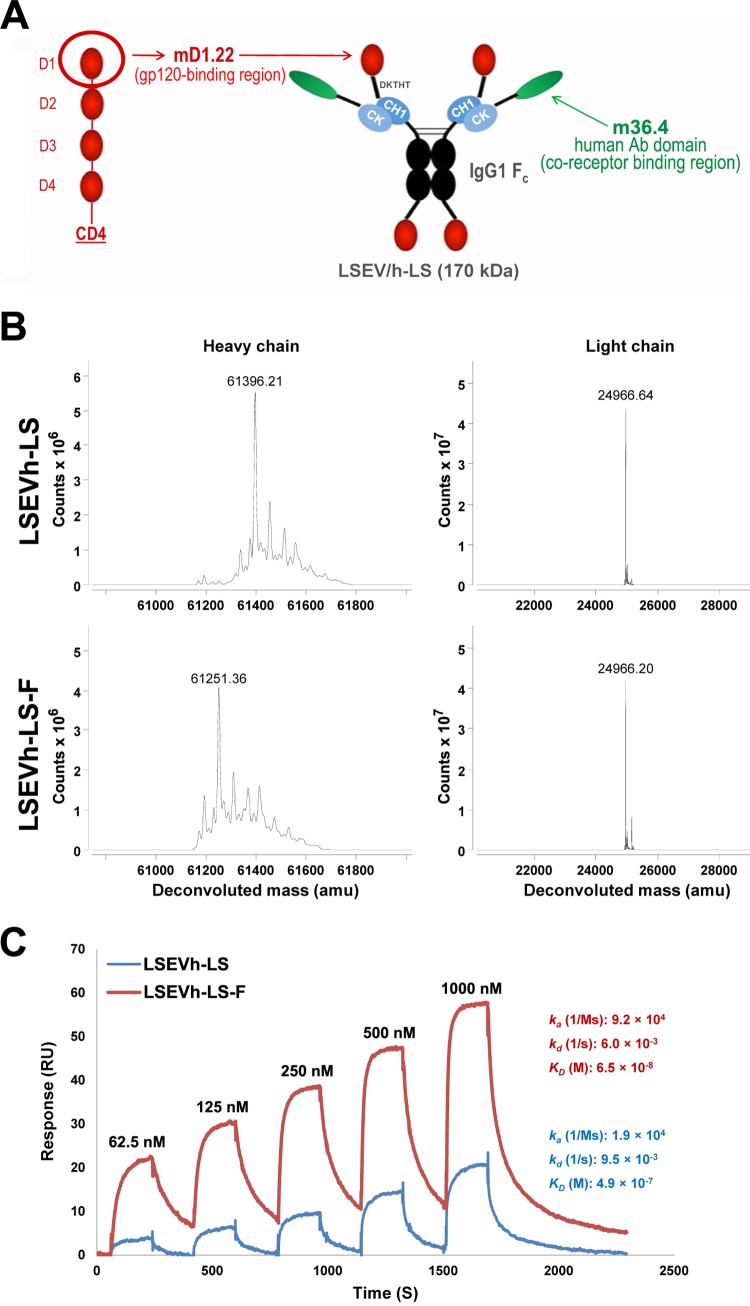
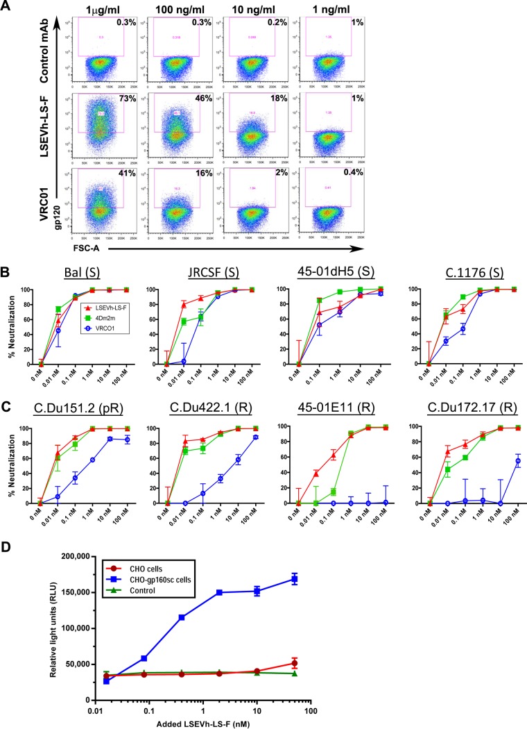
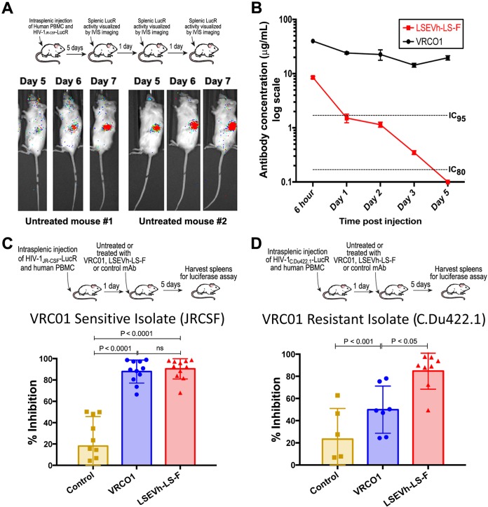
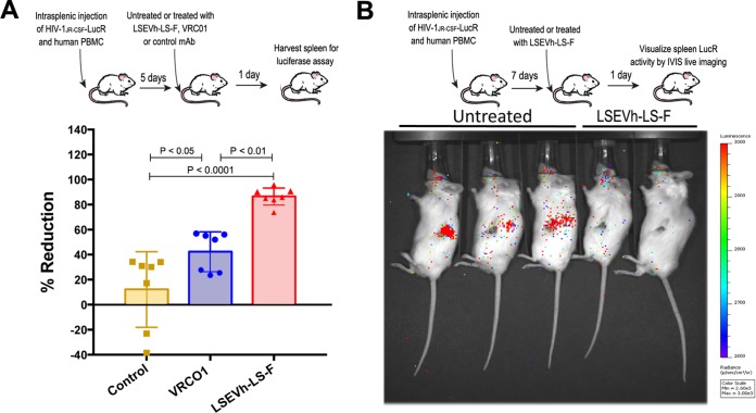
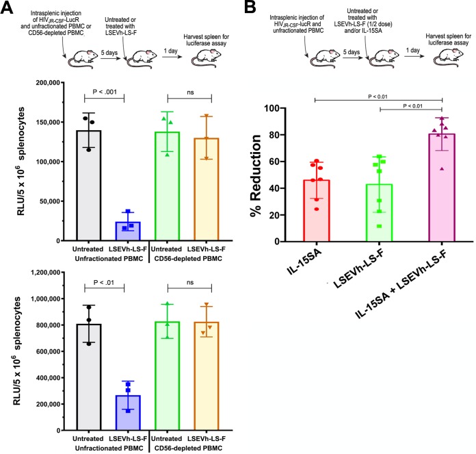
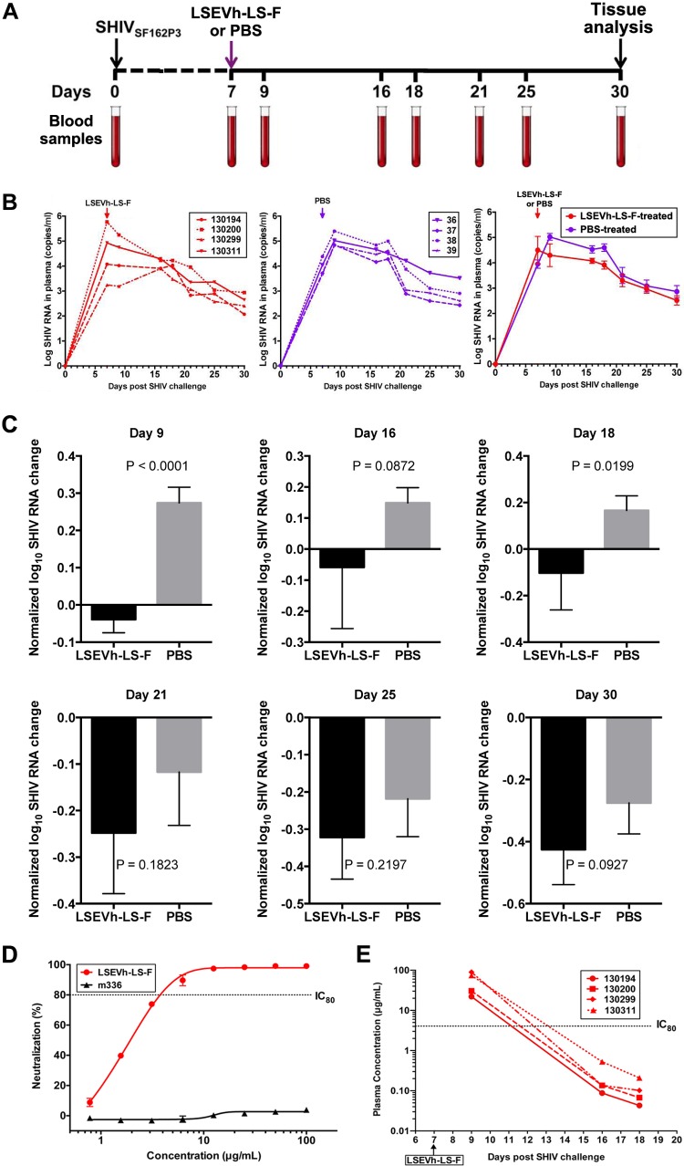
Antibodies bound to human immunodeficiency virus type 1 (HIV-1) envelope protein expressed by infected cells mobilize antibody-dependent cellular cytotoxicity (ADCC) to eliminate the HIV-1-infected cells and thereby suppress HIV-1 infection and delay disease progression. Studies treating HIV-1-infected individuals with latency reactivation agents to reduce their latent HIV-1 reservoirs indicated that their HIV-1-specific immune responses were insufficient to effectively eliminate the reactivated latent HIV-1-infected T cells. Mobilization of ADCC may facilitate elimination of reactivated latent HIV-1-infected cells to deplete the HIV-1 reservoir and contribute to a functional HIV-1 cure. The most effective antibodies for controlling and eradicating HIV-1 infection would likely have the dual capacities of potently neutralizing a broad range of HIV-1 isolates and effectively mobilizing HIV-1-specific ADCC to eliminate HIV-1-infected cells. For this purpose, we constructed LSEVh-LS-F, a broadly neutralizing, defucosylated hexavalent fusion protein specific for both the CD4 and coreceptor gp120-binding sites. LSEVh-LS-F potently inhibited in vivo HIV-1 and simian-human immunodeficiency virus (SHIV) infection in humanized mouse and macaque models, respectively, including in vivo neutralization of HIV-1 strains resistant to the broadly neutralizing antibodies VRC01 and 3BNC117. We developed a novel humanized mouse model to evaluate in vivo human NK cell-mediated elimination of HIV-1-infected cells by ADCC and utilized it to demonstrate that LSEVh-LS-F rapidly mobilized NK cells to eliminate >80% of HIV-1-infected cells in vivo 1 day after its administration. The capacity of LSEVh-LS-F to eliminate HIV-1-infected cells via ADCC combined with its broad neutralization activity supports its potential use as an immunotherapeutic agent to eliminate reactivated latent cells and deplete the HIV-1 reservoir.IMPORTANCE Mobilization of antibody-dependent cellular cytotoxicity (ADCC) to eliminate reactivated latent HIV-1-infected cells is a strategy which may contribute to depleting the HIV-1 reservoir and achieving a functional HIV-1 cure. To more effectively mobilize ADCC, we designed and constructed LSEVh-LS-F, a broadly neutralizing, defucosylated hexavalent fusion protein specific for both the CD4 and coreceptor gp120-binding sites. LSEVh-LS-F potently inhibited in vivo HIV-1 and SHIV infection in humanized mouse and macaque models, respectively, including in vivo neutralization of an HIV-1 strain resistant to the broadly neutralizing antibodies VRC01 and 3BNC117. Using a novel humanized mouse model, we demonstrated that LSEVh-LS-F rapidly mobilized NK cells to eliminate >80% of HIV-1-infected cells in vivo 1 day after its administration. The capacity of LSEVh-LS-F to eliminate HIV-1-infected cells via ADCC combined with its broad neutralization activity supports its potential use as an immunotherapeutic agent to eliminate reactivated latent cells and deplete the HIV-1 reservoir.

Keywords: NK cell; human immunodeficiency virus.


Figures

FIG 1
FIG 1
Structure, mass spectrometry analysis, and binding kinetics. (A) Schematic representation of LSEVh-LS expressing mD1.22 and m36.4. IgG1 Fc, human IgG1 crystallizable fragment; CH1, human IgG1 heavy-chain constant region 1; CK, human antibody kappa light-chain constant region; DKTHT, linker sequence derived from the human IgG1 hinge. The calculated molecular mass is shown in parentheses. (B) High-resolution mass spectrometry analysis of LSEVh-LS produced in 293-F cells, which was designated LSEVh-LS, and LSEVh-LS produced in GFT gene knockout CHOF6 cells, which was designated LSEVh-LS-F. Mass spectra were shown, with deconvoluted mass for the major peak indicated at the top. G0, Fc oligosaccharides without galactose and fucose; G0F, Fc oligosaccharides without galactose but with fucose. (C) Binding kinetics of LSEVh-LS and LSEVh-LS-F with recombinant human FcγRIIIa as measured by SPR. SPR analysis was performed on Biacore X100 by using a single-cycle approach according to the manufacturer's instructions. Analytes were tested at 1000, 500, 250, 125, and 62.5 nM concentrations. The kinetic constants shown on the right were calculated from the sensograms fitted with monovalent binding model of the BiacoreX100 evaluation software 2.0. ka, association rate constant; kd, dissociation rate constant; KD, equilibrium dissociation constant.
FIG 2
FIG 2
LSEVh-LS-F and VRC01 binding to Env expressed on the surface of ACH-2 cells, in vitro neutralizing activity, and LSEVh-LS-F in vitro ADCC activity. (A) Unactivated ACH-2 cells and PMA-activated ACH2 cells were stained with the indicated concentration of biotinylated anti-Ebola virus MAb (control), VRC01, or LSEVh-LS-F, followed by incubation with streptavidin-PE and analysis by flow cytometry. (B and C) In vitro neutralization by VRC01, 4Dm2m, or LSEVh-LS-F of PBMC infection by HIV-1Env-LucR infectious molecular clones expressing Env from the indicated VRC01-sensitive (B) and VRC01-resistant (C) HIV-1 strains. Activated PBMC from healthy donors were cultured for 48 h in the presence of HIV-1 expressing the indicated Env and the indicated concentration of VRC01, 4Dm2m, or LSEVh-LS-F. Graphed data represent the percent viral neutralization by the indicated concentrations of VRC01, 4Dm2m, and LSEVh-LS-F as measured by luciferase activity and calculated in reference to the untreated infected PBMC using the formula y = [(1 − treatment RLU)/untreated infected PMBC RLU × 100)]. Each data point in the graph represents the average value of triplicates ± standard deviation (SD). (D) In vitro ADCC activity of LSEVh-LS-F. CHO or CHO-gp160SC cells were used as target cells for measuring antibody-mediated effector function as a proxy for ADCC activity of LSEVh-LS-F using as an effector cell a genetically modified Jurkat cell line expressing the human FcRγIIIa with an inducible luciferase reporter gene. Each data point in the graph represents the average value of duplicates ± SD. The control represents no added target cells.
FIG 3
FIG 3
LSEVh-LS-F treatment significantly inhibits in vivo infection with VRC01-sensitive and VRC01-resistant HIV-1 isolates. (A) In vivo HIV-1JRCSF-LucR infection was visualized using IVIS imaging on days 5, 6, and 7 after viral inoculation. Images were acquired for 3 min after substrate injection and have been corrected for background bioluminescence. Scans of two representative mice are shown. (B) Pharmacokinetics of LSEVh-LS-F and VRC01 in vivo in NSG mice. After NSG mice (n = 2) were injected with either VRC01 or LSEVh-LS-F, serum was collected 6 h, 1 day, 2 days, 3 days, and 5 days later, and the levels of antibody in each serum sample were determined using a gp140 ELISA. The results are presented on a log scale. The dotted lines on the graph indicate the LSEVh-LS-F levels that neutralize 80% (IC80, 0.17 μg) or at least 95% (IC95, 1.7 μg) of VRC01-sensitive HIV-1 in vitro. (C and D) hu-spl-PBMC-NSG mice were inoculated with HIV-1Env-LucR expressing the VRC01-susceptible Env from HIV-1 strain JR-CSF (C) or the VRC01-resistant Env from HIV-1 strain C.Du422.1 (D). The next day, groups of mice were either left untreated or treated with 0.5 mg of either LSEVh-LS-F, VRC01, or control antibody m336. Five days later, HIV-1 infection of the mice was measured by quantifying LucR activity in the splenic lysates. A dot plot graph displays the percent neutralization in each mouse treated with the indicated treatment compared to the untreated group and the group mean ± SD and represent the pooled data from 3 independent experiments using 3 different donors (C) or from 2 independent experiments using 2 different donors (D).
FIG 4
FIG 4
LSEVh-LS-F mobilizes ADCC-mediated in vivo elimination of HIV-1-infected cells. (A) Five days after hu-spl-PBMC-NSG mice were inoculated with HIV-1JRCSF-LucR, they were either left untreated or treated with 0.5 mg of LSEVh-LS-F, VRC01, or control antibody m336, and HIV-1 infection was determined by quantifying LucR activity in the splenic lysates the next day. The percent reduction for each mouse compared to the untreated group is presented as a dot plot graph with the group mean ± SD of pooled data from 2 independent experiments using 2 different donors. (B) Seven days after hu-spl-PBMC-NSG mice were inoculated with HIV-1JRCSF-LucR, they were either left untreated or treated with LSEVh-LS-F (0.5 mg). HIV-1 infection indicated by LucR activity was visualized by IVIS live imaging of bioluminescence after injection of the mice with the substrate.
FIG 5
FIG 5
LSEVh-LS-F-induced ADCC requires NK cells for elimination of HIV-1-infected cells. (A) Five days after unfractionated or NK-cell-depleted PBMC were intrasplenically injected into NSG mice (∼107 cells) together with HIV-1JRCSF-LucR, the mice were left untreated or treated with LSEVh-LS-F (0.5 mg), and 1 day later, LucR activity in the splenic lysates was quantified. Data from two independent experiments are shown as dot plots with the group mean ± SD for each experiment. (B) Five days after hu-spl-PBMC-NSG mice were intrasplenically injected with HIV-1JRCSF-LucR (1 × 10⁷ IU), mice were either left untreated or treated with LSEVh-LS-F (0.25 mg) and/or ALT-803 (0.2 mg/kg), and 1 day later, LucR activity in the splenic lysates was quantified. The percent suppression of infection for each mouse compared to the untreated group is shown as a dot plot with mean ± SD for the group. The graph represents pooled data from 2 independent experiments using one donor.
FIG 6
FIG 6
Effect of LSEVh-LS-F treatment on plasma SHIV loads. (A) Experimental treatment and analysis scheme. (B) Eight rhesus macaques were challenged intravenously with SHIV-1SF162P3 and treated on day 7 with either a single infusion of LSEVh-LS-F (n = 4) or PBS (n = 4). Plasma SHIV loads from individual LSEVh-LS-F-treated macaques (left panel) and individual PBS-treated macaques (middle panel) and the average for the four macaques in the LSEVh-LS-F-treated and PBS-treated groups (right panel) are shown. (C) Analysis with normalized values of the data shown in panel B was performed to decrease the effect of individual macaque variation. Evaluation of the difference in plasma SHIV loads between the treatment and control groups was analyzed by the unpaired t test for different days after treatment. The decrease of virus RNA at day 9 for LSEVh-LS-F-treated compared to PBS-treated macaques is highly statistically significant (P = 0.0001). (D) Dose-response in vitro neutralization of SHIVSF162P3 by LSEVh-LS-F assayed in TZM-bl cells. An irrelevant monoclonal antibody, m336, was used as the negative control. (E) Concentration of LSEVh-LS-F after a single infusion of LSEVh-LS-F as a function of time for the individual treated macaques. The limit of detection is about 0.1 μg/ml, and after day 18, the LSEVh-LS-F concentration can be assumed to be undetectable.
FIG 7
FIG 7
Effect of LSEVh-LS-F treatment on SHIV infection of tissues. Quantification of SHIV RNA in mucosal tissues (A) and lymph nodes, brain, and testes/ovaries (B) is shown. The number of SHIV RNA copies/μg of the indicated tissue in the LSEVh-LS-F-treated and untreated macaques was determined by RT-qPCR at day 30 and is shown for each macaque in a dot plot graph with the mean ± SD for the group.

References

    1. Walker LM, Huber M, Doores KJ, Falkowska E, Pejchal R, Julien JP, Wang SK, Ramos A, Chan-Hui PY, Moyle M, Mitcham JL, Hammond PW, Olsen OA, Phung P, Fling S, Wong CH, Phogat S, Wrin T, Simek MD, Koff WC, Wilson IA, Burton DR, Poignard P. 2011. Broad neutralization coverage of HIV by multiple highly potent antibodies. Nature 477:466–470. doi:10.1038/nature10373. - DOI - PMC - PubMed
    1. Euler Z, Alter G. 2015. Exploring the potential of monoclonal antibody therapeutics for HIV-1 eradication. AIDS Res Hum Retroviruses 31:13–24. doi:10.1089/aid.2014.0235. - DOI - PMC - PubMed
    1. Lee WS, Parsons MS, Kent SJ, Lichtfuss M. 2015. Can HIV-1-specific ADCC assist the clearance of reactivated latently infected cells? Front Immunol 6:265. doi:10.3389/fimmu.2015.00265. - DOI - PMC - PubMed
    1. Bruel T, Guivel-Benhassine F, Amraoui S, Malbec M, Richard L, Bourdic K, Donahue DA, Lorin V, Casartelli N, Noel N, Lambotte O, Mouquet H, Schwartz O. 2016. Elimination of HIV-1-infected cells by broadly neutralizing antibodies. Nat Commun 7:10844. doi:10.1038/ncomms10844. - DOI - PMC - PubMed
    1. Horwitz JA, Halper-Stromberg A, Mouquet H, Gitlin AD, Tretiakova A, Eisenreich TR, Malbec M, Gravemann S, Billerbeck E, Dorner M, Buning H, Schwartz O, Knops E, Kaiser R, Seaman MS, Wilson JM, Rice CM, Ploss A, Bjorkman PJ, Klein F, Nussenzweig MC. 2013. HIV-1 suppression and durable control by combining single broadly neutralizing antibodies and antiretroviral drugs in humanized mice. Proc Natl Acad Sci U S A 110:16538–16543. doi:10.1073/pnas.1315295110. - DOI - PMC - PubMed

Publication types

MeSH terms