. 2017 Oct 13;91(21):e00855-17.
doi: 10.1128/JVI.00855-17. Print 2017 Nov 1.

Recombinant Origins of Pathogenic and Nonpathogenic Mouse Gammaretroviruses with Polytropic Host Range

Affiliations

Recombinant Origins of Pathogenic and Nonpathogenic Mouse Gammaretroviruses with Polytropic Host Range

Devinka Bamunusinghe et al. J Virol. .

Abstract

Ecotropic, xenotropic, and polytropic mouse leukemia viruses (E-, X-, and P-MLVs) exist in mice as infectious viruses and endogenous retroviruses (ERVs) inserted into mouse chromosomes. All three MLV subgroups are linked to leukemogenesis, which involves generation of recombinants with polytropic host range. Although P-MLVs are deemed to be the proximal agents of disease induction, few biologically characterized infectious P-MLVs have been sequenced for comparative analysis. We analyzed the complete genomes of 16 naturally occurring infectious P-MLVs, 12 of which were typed for pathogenic potential. We sought to identify ERV progenitors, recombinational hot spots, and segments that are always replaced, never replaced, or linked to pathogenesis or host range. Each P-MLV has an E-MLV backbone with P- or X-ERV replacements that together cover 100% of the recombinant genomes, with different substitution patterns for X- and P-ERVs. Two segments are always replaced, both coding for envelope (Env) protein segments: the N terminus of the surface subunit and the cytoplasmic tail R peptide. Viral gag gene replacements are influenced by host restriction genes Fv1 and Apobec3 Pathogenic potential maps to the env transmembrane subunit segment encoding the N-heptad repeat (HR1). Molecular dynamics simulations identified three novel interdomain salt bridges in the lymphomagenic virus HR1 that could affect structural stability, entry or sensitivity to host immune responses. The long terminal repeats of lymphomagenic P-MLVs are differentially altered by recombinations, duplications, or mutations. This analysis of the naturally occurring, sometimes pathogenic P-MLV recombinants defines the limits and extent of intersubgroup recombination and identifies specific sequence changes linked to pathogenesis and host interactions.IMPORTANCE During virus-induced leukemogenesis, ecotropic mouse leukemia viruses (MLVs) recombine with nonecotropic endogenous retroviruses (ERVs) to produce polytropic MLVs (P-MLVs). Analysis of 16 P-MLV genomes identified two segments consistently replaced: one at the envelope N terminus that alters receptor choice and one in the R peptide at the envelope C terminus, which is removed during virus assembly. Genome-wide analysis shows that nonecotropic replacements in the progenitor ecotropic MLV genome are more extensive than previously appreciated, covering 100% of the genome; contributions from xenotropic and polytropic ERVs differentially alter the regions responsible for receptor determination or subject to APOBEC3 and Fv1 restriction. All pathogenic viruses had modifications in the regulatory elements in their long terminal repeats and differed in a helical segment of envelope involved in entry and targeted by the host immune system. Virus-induced leukemogenesis thus involves generation of complex recombinants, and specific replacements are linked to pathogenesis and host restrictions.

Keywords: origins of infectious gammaretroviruses; pathogenic mouse gammaretroviruses; polytropic mouse gammaretroviruses; recombinant retroviruses; retroviral N-heptad repeat.


Figures

FIG 1
FIG 1
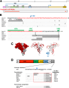
Alignments of 16 P-MLVs showing segments of homology to E-, P-, and X-MLV ERVs. The diagram at the top indicates the positions of LTRs and coding regions. Individual isolates are identified on the left. E-MLV-related segments are indicated in black, Pmvs are indicated in red/pink, and Xmv are indicated in greens, and boxes indicate segments where the identity to known ERVs is <96%. Numbers at the junction sites are the last matching nucleotide in that segment. At the bottom are shown the total genome replacements by Pmv and Xmv ERVs, and the total combined nonecotropic replacements.
FIG 2
FIG 2
Alignments of 16 P-MLVs showing segments of homology with E-, P-, and X-MLV ERVs. A diagram at the top indicates positions of LTRs and coding regions. Individual isolates are named on the left. Segmental subgroup relationships are color coded. Numbers below the line drawing of each P-MLV represent the percent identity to the Emv progenitor, and numbers above are the percent identities to X- or P-ERVs of segments replaced by recombination.
FIG 3
FIG 3
Three clusters of recombination breakpoints in and around env. Red boxes identify segments of template switching between the E-MLV and Pmv genomes. Twelve of the sixteen viruses have breakpoints in segment A, and segments B and C contain breakpoints for four and eight viruses, respectively. Viruses using breakpoint regions are named above the boxes. Segments with crossover sites for two or more viruses are highlighted in yellow.
FIG 4
FIG 4
Two Pmv env substitutions in all 16 P-MLVs. (A) Diagram of the MLV Env showing the surface (SU) and transmembrane (TM) subunits, leader region (pink), the three variable domains (VRA, VRB, and VRC), the RBD, the proline-rich region (PRR), and the cytoplasmic tail (CT). Two red bars indicate the minimum common Pmv replacement in all 16 viruses. (B) Protein sequences of the Env leader and SU N terminus for six P-MLVs with recombination breakpoints in this region are compared with the Pmv13 ERV. The minimum shared P-MLV replacement sequence is shown in red. Gray highlights identify homologous regions involved in recombination that are within the displayed sequence. (C) The Pmv Env replacement covers much of the RBD apex as shown in surface and ribbon representations of the RBD structure based on (45). (D) Protein sequences of the CT of 7 P-MLVs with CT recombination breakpoints compared to parental MLV ERVs. Gray highlights identify regions of recombination that lie within the indicated sequence, the blue arrow marks the R-peptide cleavage site, and a red box identifies the common replacement.
FIG 5
FIG 5
P-MLV substitutions that alter the target of the Fv1 restriction factor. Three P-MLVs have Xmv (green) or Pmv (red) substitutions in the Fv1 target region in capsid. P-MLV sequences are compared to prototype MLVs with Fv1 N-tropism (AKV) and B-tropism (WN1802B) and with their likely ERV progenitors. Numbers identify residues that affect sensitivity to Fv1; residue 110 is the major determinant of N- or B-tropism.
FIG 6
FIG 6
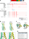
Substitution of the HR1 of TMenv is linked to pathogenesis. (A) Sequence variation in 12 P-MLVs within the minimum Env segment that distinguishes all lymphomagenic from all nonlymphomagenic viruses. At the top is shown the domain structure of the TM Env. Sequences are compared to AKV E-MLV and Pmv13. Marked are eight substitutions that alter charge or that change the key a or d heptad repeat sites. Blue arrows mark residues involved in altered salt bridge formation. (B) Predicted structures of the six-helix bundles of the lymphomagenic virus AKR-L4, and the nonlymphomagenic virus Akv2-M66 based on the structure of MPMV (PDB 4JF3), models used for molecular dynamics simulations (see Movie S1 in the supplemental material). Key residues are labeled, and the chloride ion that stabilizes the structure is represented by a green ball. An enlargement of the predicted HR1-HR2 interfaces shows key residues that are likely to form salt bridges. (C) Models of the two predicted trimers of heterodimers. Residues that distinguish the two are labeled, and the chloride ion is shown as a green ball. (D) Predicted frequency of formation of five HR1-HR2 interdomain salt bridges in the lymphomagenic and nonlymphomagenic P-MLVs.

References

    1. Kozak CA. 2015. Origins of the endogenous and infectious laboratory mouse gammaretroviruses. Viruses 7:1–26. doi:10.3390/v7010001. - DOI - PMC - PubMed
    1. Jenkins NA, Copeland NG, Taylor BA, Lee BK. 1982. Organization, distribution, and stability of endogenous ecotropic murine leukemia virus DNA sequences in chromosomes of Mus musculus. J Virol 43:26–36. - PMC - PubMed
    1. O'Neill RR, Khan AS, Hoggan MD, Hartley JW, Martin MA, Repaske R. 1986. Specific hybridization probes demonstrate fewer xenotropic than mink cell focus-forming murine leukemia virus env-related sequences in DNAs from inbred laboratory mice. J Virol 58:359–366. - PMC - PubMed
    1. Fischinger PJ, Nomura S, Bolognesi DP. 1975. A novel murine oncornavirus with dual eco- and xenotropic properties. Proc Natl Acad Sci U S A 72:5150–5155. doi:10.1073/pnas.72.12.5150. - DOI - PMC - PubMed
    1. Hartley JW, Wolford NK, Old LJ, Rowe WP. 1977. New class of murine leukemia-virus associated with development of spontaneous lymphomas. Proc Natl Acad Sci U S A 74:789–792. doi:10.1073/pnas.74.2.789. - DOI - PMC - PubMed

Publication types

MeSH terms

LinkOut - more resources