. 2017 Nov 1;313(5):F1097-F1105.
doi: 10.1152/ajprenal.00261.2017. Epub 2017 Aug 9.

Vasa recta pericyte density is negatively associated with vascular congestion in the renal medulla following ischemia reperfusion in rats

Affiliations

Vasa recta pericyte density is negatively associated with vascular congestion in the renal medulla following ischemia reperfusion in rats

G Ryan Crislip et al. Am J Physiol Renal Physiol. .

Abstract

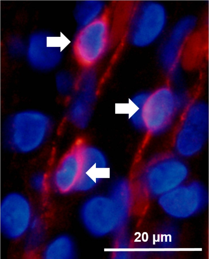
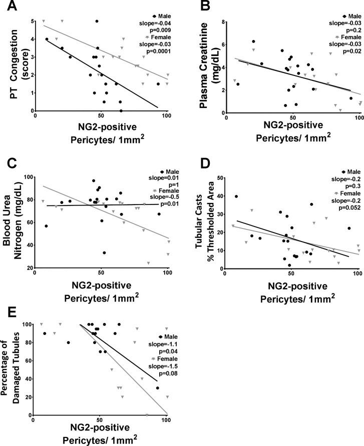
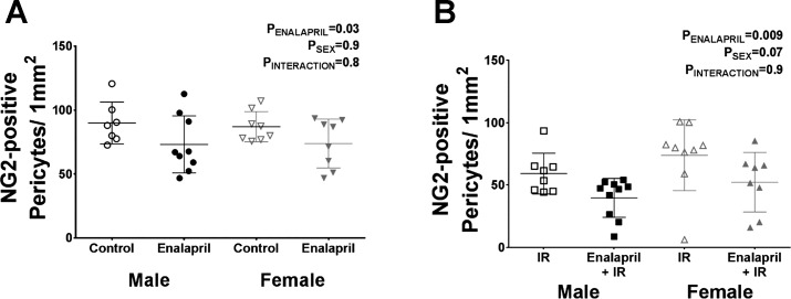
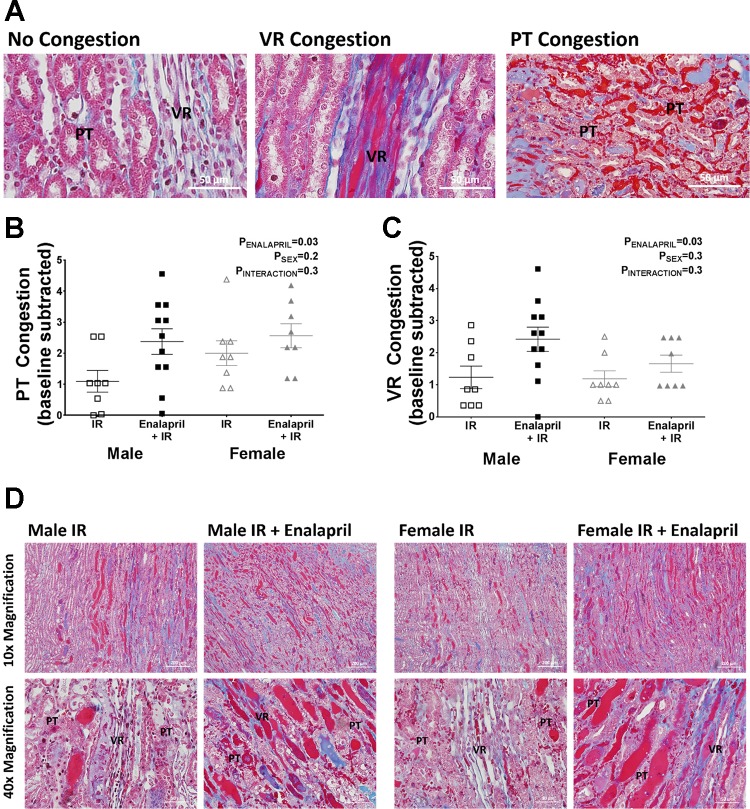
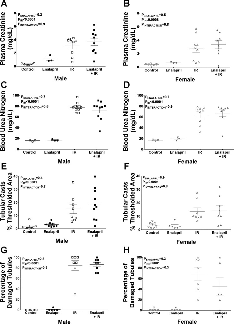
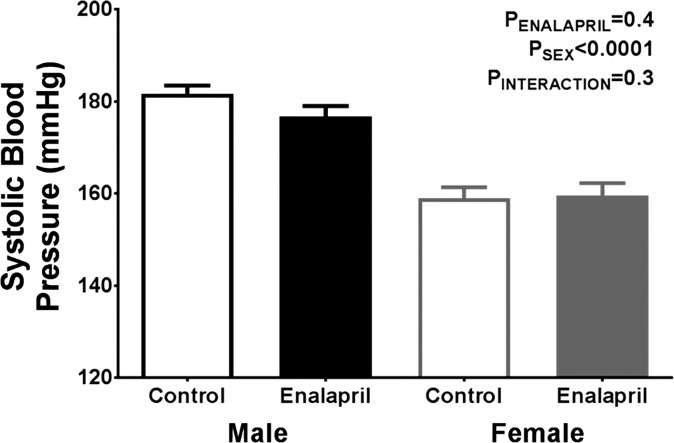
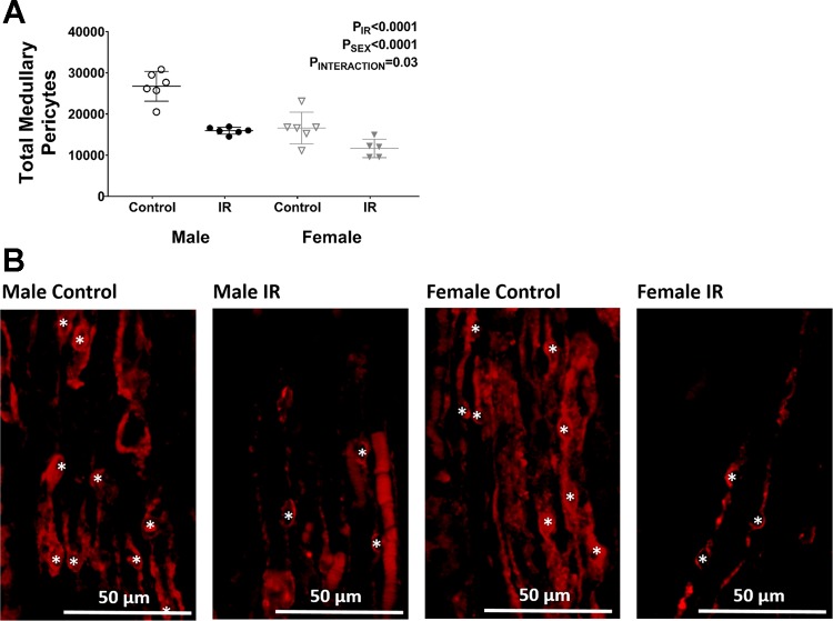
Recent evidence suggests that a greater density of pericytes in renal cadaveric allografts is associated with better recovery following transplant. The physiological mechanism(s) through which pericyte density may be beneficial is not well understood. The goal of this study was to test the hypothesis that lower medullary pericyte density is associated with greater renal injury following ischemia reperfusion (IR) in a rat model, providing a basis for future studies to better understand pericytes in a pathological environment. To test our hypothesis, we determined the association between medullary pericyte density and renal injury in spontaneously hypertensive rats (SHR) following 45 min of warm bilateral IR. We found that there was a significant negative relationship between pericyte density and plasma creatinine (slope = -0.03, P = 0.02) and blood urea nitrogen (slope = -0.5, P = 0.01) in female but not male SHR. Pericyte density was negatively associated with medullary peritubular capillary (PT) congestion in both sexes following IR (male: slope = -0.04, P = 0.009; female: slope = -0.03, P = 0.0001). To further test this relationship, we used a previously reported method to reduce pericyte density in SHR. Medullary erythrocyte congestion in vasa recta (VR) and PT significantly increased following IR in both sexes when pericyte density was pharmacologically decreased (VR: P = 0.03; PT: P = 0.03). Our data support the hypothesis that pericyte density is negatively associated with the development of IR injury in SHR, which may be mediated by erythrocyte congestion in the medullary vasculature.

Keywords: erythrocyte trapping; ischemia reperfusion injury; sex difference; spontaneously hypertensive rats.


Figures

Fig. 1.
Fig. 1.
Representative image of fluorescently labeled neural glial 2 (NG2)-positive pericyte cell bodies overlapped with DAPI staining of nuclei, which was used for the quantification of pericytes. White arrows point to the three pericyte cells that are visible; image was taken from an ischemia-reperfusion (IR) male. Note the width of the vessels in relation to diameter of the light-red-stained red blood cells (RBCs) that can be seen in the image.
Fig. 2.
Fig. 2.
Correlation of renal injury markers and pericyte density determined by NG2 immunofluorescence. Data from each individual animal that underwent IR surgery are shown; males are represented by black circles and females by gray triangles. The markers that were compared with pericyte density include peritubular capillary (PT) congestion (A), plasma creatinine (B), blood urea nitrogen (C), percent area with tubular casts (D), and percentage of damaged tubules (E). Regression slopes and P values are shown; n = 13–18.
Fig. 3.
Fig. 3.
Effect of transient enalapril treatment on pericyte density determined by NG2 immunofluorescence. Pericyte density was determined in vehicle- and enalapril-treated rats (A) and in treated rats following IR (B) to confirm that enalapril treatment decreases pericyte density. Values are expressed as means ± SE; n = 7–10.
Fig. 4.
Fig. 4.
Effect of transient enalapril treatment on medullary vascular congestion following IR. Incidence of RBC congestion was assessed in peritubular (PT) and vasa recta (VR) capillaries post-IR independently of one another, where a score of 0 indicates all visible vessels are open and 5 indicates all visible vessels are congested. PT capillaries were determined as vessels found between tubular structures and VR as bundles of long vessels. A: representative images show the distinction between these vessels and an example of congested and uncongested PT and VR. Scatterplots show the respective congestion scores from each individual animal in PT (B) and VR (C). Baseline levels for each respective group of rats were subtracted to eliminate any baseline congestion that occurred in animals with no surgery. D: representative images used for scoring the kidneys from each treatment group that had IR surgery are shown. RBCs and protein casts are stained in red. Values are expressed as means ± SE; n = 8–11 for scatterplots.
Fig. 5.
Fig. 5.
Effect of transient enalapril treatment on renal injury in adulthood. Renal injury markers were measured to determine whether enalapril treatment had an effect on IR-induced injury in either sex. Plasma creatinine (male: A; female: B), blood urea nitrogen (BUN; male: C; female: D), tubular cast area (male: E; female: F), and the percentage of tubules damaged (male: G; female: H) are shown. Values are expressed as means ± SE; n = 3 for rats without IR due to limited number of samples for plasma creatinine and BUN assays; n = 5–10 for the rest of the groups.
Fig. 6.
Fig. 6.
Effect of transient enalapril on blood pressure in adulthood. Tail cuff method was used to measure systolic blood pressure in vehicle and enalapril-treated rats at 13 wk of age. Values are expressed as means ± SE; n = 8–13.
Fig. 7.
Fig. 7.
Quantification of pericyte number. A: total pericyte number was determined in untreated rats before and after IR by multiplying the volume of the outer medulla and pericyte density determined by neural glial 2 (NG2) immunofluorescence. B: representative images show the decrease in pericyte density following IR in both sexes. Asterisks indicate NG2-positive pericyte cell bodies used for quantification. Values are expressed as means ± SE; n = 5 or 6.

Comment in

  • Recent advances in sex differences in kidney function.
    Layton AT, Sullivan JC. Layton AT, et al. Am J Physiol Renal Physiol. 2019 Feb 1;316(2):F328-F331. doi: 10.1152/ajprenal.00584.2018. Epub 2018 Dec 19. Am J Physiol Renal Physiol. 2019. PMID: 30565997 Free PMC article. No abstract available.

References

    1. Adams MA, Bobik A, Korner PI. Enalapril can prevent vascular amplifier development in spontaneously hypertensive rats. Hypertension 16: 252–260, 1990. [Erratum in Hypertension 17: 262, 1991.] doi: 10.1161/01.HYP.16.3.252. - DOI - PubMed
    1. Armulik A, Genové G, Betsholtz C. Pericytes: developmental, physiological, and pathological perspectives, problems, and promises. Dev Cell 21: 193–215, 2011. doi: 10.1016/j.devcel.2011.07.001. - DOI - PubMed
    1. Baumann M, Janssen BJ, Hermans JJ, Peutz-Kootstra C, Witzke O, Smits JF, Struijker Boudier HA. Transient AT1 receptor-inhibition in prehypertensive spontaneously hypertensive rats results in maintained cardiac protection until advanced age. J Hypertens 25: 207–215, 2007. doi: 10.1097/HJH.0b013e3280102bff. - DOI - PubMed
    1. Baumann M, Janssen BJ, Rob Hermans JJ, Bartholome R, Smits JF, Struijker Boudier HA. Renal medullary effects of transient prehypertensive treatment in young spontaneously hypertensive rats. Acta Physiol (Oxf) 196: 231–237, 2009. doi: 10.1111/j.1748-1716.2008.01916.x. - DOI - PubMed
    1. Bergers G, Song S. The role of pericytes in blood-vessel formation and maintenance. Neuro-oncol 7: 452–464, 2005. doi: 10.1215/S1152851705000232. - DOI - PMC - PubMed

LinkOut - more resources