. 2017 Dec;28(12):3688-3698.
doi: 10.1681/ASN.2016101074. Epub 2017 Aug 9.

Tubulointerstitial Nephritis with IgM-Positive Plasma Cells

Affiliations

Tubulointerstitial Nephritis with IgM-Positive Plasma Cells

Naoki Takahashi et al. J Am Soc Nephrol. 2017 Dec.

Abstract

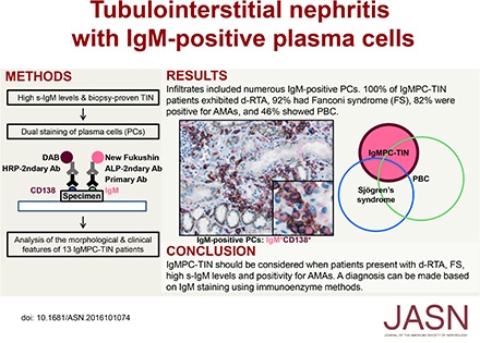
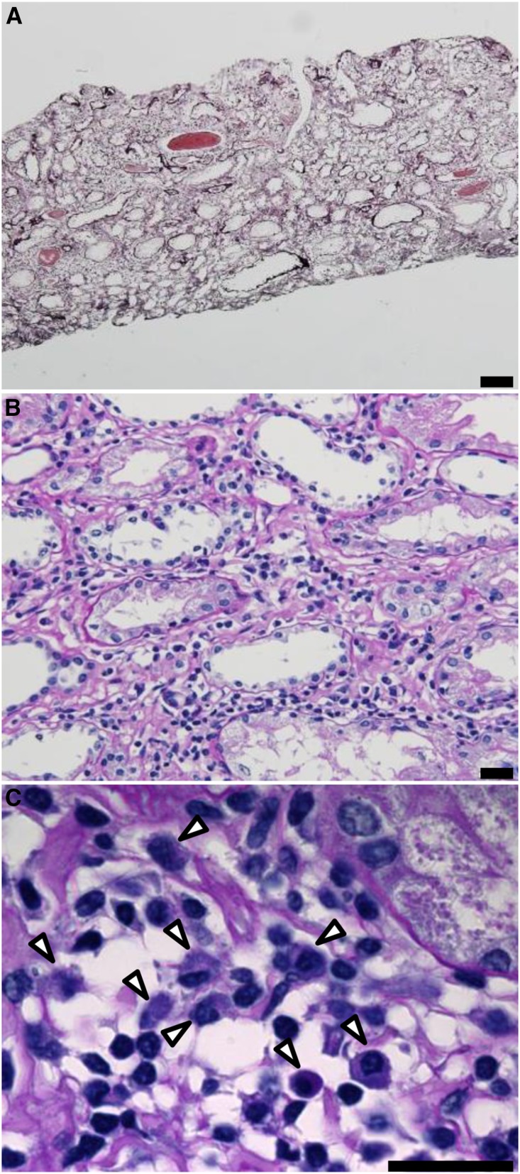
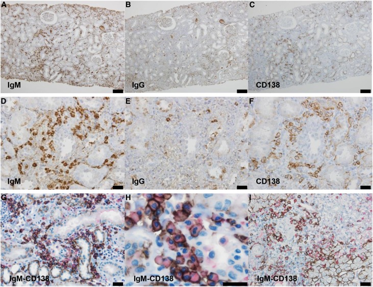
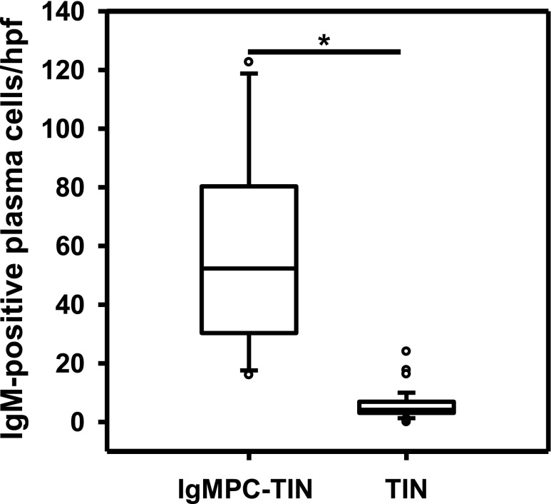
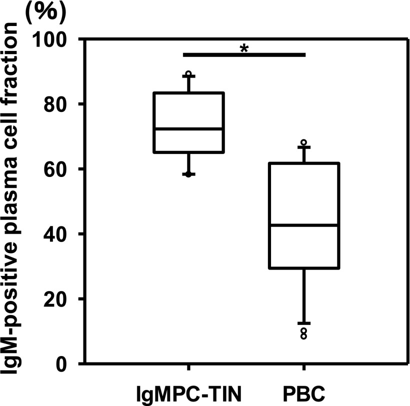
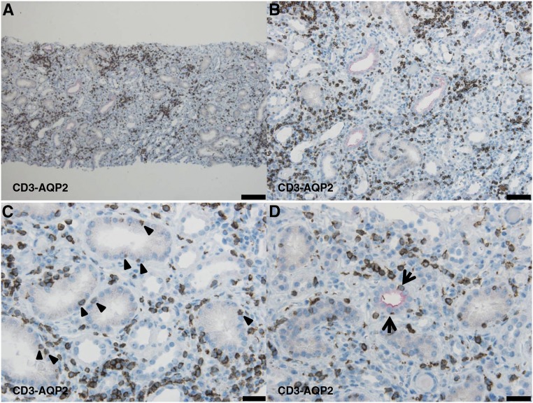
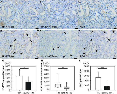
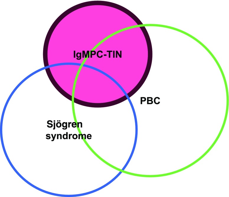
Infiltration by IgG-positive plasma cells is a common finding in tubulointerstitial nephritis. Indeed, it has been thought that CD138-positive mature plasma cells secrete mainly IgG, and the occurrence of tubulointerstitial nephritis with CD138-positive plasma cells secreting IgM has rarely been reported. Routine immunofluorescence of fresh frozen sections is considered the gold standard for detection of immune deposits. However, the immunoenzyme method with formalin-fixed, paraffin-embedded sections is superior for detecting IgM- or IgG-positive cells within the renal interstitium, thus histologic variants may often go undetected. We recently discovered a case of tubulointerstitial nephritis showing IgM-positive plasma cell accumulation within the interstitium. To further explore the morphologic and clinical features of such cases, we performed a nationwide search for patients with biopsy-proven tubulointerstitial nephritis and high serum IgM levels. We identified 13 patients with tubulointerstitial nephritis and IgM-positive plasma cell infiltration confirmed with the immunoenzyme method. The clinical findings for these patients included a high prevalence of distal renal tubular acidosis (100%), Fanconi syndrome (92%), and anti-mitochondrial antibodies (82%). The pathologic findings were interstitial nephritis with diffusely distributed CD3-positive T lymphocytes and colocalized IgM-positive plasma cells, as well as tubulitis with CD3-positive T lymphocytes in the proximal tubules and collecting ducts. Additionally, levels of H+-ATPase, H+, K+-ATPase, and the HCO3--Cl- anion exchanger were markedly decreased in the collecting ducts. We propose to designate this group of cases, which have a common histologic and clinical form, as IgM-positive plasma cell-tubulointerstitial nephritis.

Keywords: Fanconi syndrome; IgM; plasma cell; renal tubular acidosis (RTA); tubulointerstitial nephritis (TIN).


Figures

None
Graphical abstract
Figure 1.
Figure 1.
Light microscopy from IgMPC-TIN patients showed interstitial nephritis and tubulitis. (A) Representative photomicrograph showing interstitial nephritis with an area of tubular atrophy and mild fibrosis (patient 1). However, storiform fibrosis, eosinophil infiltration, and obliterative phlebitis, which are characteristic findings of IgG4-related disease, were not observed (periodic acid–methenamine silver staining). Bar = 100 µm. (B) Higher magnification image showing infiltration by lymphocytes and plasma cells into the interstitium (periodic acid–Schiff staining). Bar = 20 µm. (C) Highest magnification image showing infiltration by lymphocytes and plasma cells (arrowheads) into the interstitium. Bar = 20 µm.
Figure 2.
Figure 2.
Immunohistochemical staining revealed the presence of many IgM-positive plasma cells within the renal interstitium. Representative images of renal (A–H) and hepatologic (I) immunohistochemical staining of infiltrating cells in patients 3 (A–F) and 1 (G and H) with IgMPC-TIN and a patient with PBC (I). Light photomicrographs show IgM-positive cells (A and D), IgG-positive cells (B and E), and CD138-positive plasma cells (C and F) within the renal interstitium. The interstitial cellular infiltrates were composed of diffusely spread IgM-positive cells. Many IgM-positive cells infiltrating the renal interstitium (G and H) and portal tract (I) were also CD138-positive (red: IgM; brown: CD138). (A–C): Bar =100 µm; (D–G, and I): bar =50 µm; (H): bar =20 µm.
Figure 3.
Figure 3.
The averaged number of infiltrating IgM-positive plasma cells per high-power field of renal interstitium from patients (n=13) with IgMPC-TIN was significantly higher than from patients (n=44) with other forms of TIN chosen as controls for staining. *P<0.001.
Figure 4.
Figure 4.
The IgM-positive plasma cell fraction in the renal interstitium of patients with IgMPC-TIN (n=13) was significantly higher than in the portal tract of patients with PBC (n=28). The IgM-positive plasma cell fraction among CD138-positive cells was calculated as the percentage of dual-positive IgM-CD138 cells among the total CD138-positive cells. *P<0.001.
Figure 5.
Figure 5.
The dual staining of CD3 and AQP2 shows tubulitis in the proximal tubules and collecting ducts. Representative light photomicrographs (patient 13) show that infiltrating CD3-positive T lymphocytes were diffusely distributed in the renal interstitium (A and B), among proximal tubular epithelial cells (proximal tubulitis) (arrowheads) (C), and among collecting duct epithelial cells (collecting duct tubulitis) (arrow) (D) (red: AQP2, brown: CD3). (A): Bar = 100 μm; (B): bar = 50 μm; (C and D): bar = 20 μm.
Figure 6.
Figure 6.
The levels of proton pumps and AE-1 in CCDs were significantly lower in patients with IgMPC-TIN than in control patients with TIN without d-RTA. The photomicrographs show the greatly diminished levels or absence of H+-ATPase (A), H+, K+-ATPase (B), and AE-1 (C) in CCDs from patient 13 with IgMPC-TIN, and the mildly decreased levels (arrowheads) of H+-ATPase (D), H+, K+-ATPase (E), and AE-1 (F) in CCDs from a patient with TIN without d-RTA. Bar = 20 μm. In quantitative image analysis of renal immunohistochemical staining, the levels of H+-ATPase (G), H+, K+-ATPase (H), and AE-1 (I) in CCDs from patients with IgMPC-TIN (n=13) were significantly lower than those in CCDs from control TIN patients (n=10) without d-RTA. *P<0.05; **P<0.01; ***P<0.001.
Figure 7.
Figure 7.
Relations among IgMPC-TIN (red) and overlapping diseases. Only 46% of patients with IgMPC-TIN satisfied the criteria for PBC, whereas 31% satisfied the criteria for Sjӧgren syndrome.

Similar articles

Cited by

References

    1. Churg J, Cotran R, Sinniah R, Sakaguchi H, Sobin L: Renal Disease: Classification and Atlas of Tubulo-Interstitial Diseases, Tokyo, Igaku-Shoin, 1985
    1. Colvin R, Fang L: Interstitial nephritis. In: Renal Pathology with Clinical and Functional Correlations, edited by Tisher C, Brenner B, Philadelphia, JB Lippincott, 1994, pp 723–768
    1. Saeki T, Nishi S, Imai N, Ito T, Yamazaki H, Kawano M, Yamamoto M, Takahashi H, Matsui S, Nakada S, Origuchi T, Hirabayashi A, Homma N, Tsubata Y, Takata T, Wada Y, Saito A, Fukase S, Ishioka K, Miyazaki K, Masaki Y, Umehara H, Sugai S, Narita I: Clinicopathological characteristics of patients with IgG4-related tubulointerstitial nephritis. Kidney Int 78: 1016–1023, 2010 - PubMed
    1. Saeki T, Kawano M: IgG4-related kidney disease. Kidney Int 85: 251–257, 2014 - PubMed
    1. Takahashi N, Kimura H, Kawajiri Y, Mikami D, Yamamoto C, Kasuno K, Imai N, Kuroda T, Nishi S, Yamamoto M, Yoshida H: Tubulointerstitial nephritis with IgM-positive plasmacytoid large lymphocyte infiltration in a patient with primary biliary cirrhosis and Sjögren’s syndrome. Clin Nephrol 74: 74–80, 2010 - PubMed

Substances

LinkOut - more resources