. 2017 Oct 6;292(40):16616-16625.
doi: 10.1074/jbc.M117.790451. Epub 2017 Aug 9.

Both brown adipose tissue and skeletal muscle thermogenesis processes are activated during mild to severe cold adaptation in mice

Affiliations

Both brown adipose tissue and skeletal muscle thermogenesis processes are activated during mild to severe cold adaptation in mice

Naresh C Bal et al. J Biol Chem. .

Abstract

Thermogenesis is an important homeostatic mechanism essential for survival and normal physiological functions in mammals. Both brown adipose tissue (BAT) (i.e. uncoupling protein 1 (UCP1)-based) and skeletal muscle (i.e. sarcolipin (SLN)-based) thermogenesis processes play important roles in temperature homeostasis, but their relative contributions differ from small to large mammals. In this study, we investigated the functional interplay between skeletal muscle- and BAT-based thermogenesis under mild versus severe cold adaptation by employing UCP1-/- and SLN-/- mice. Interestingly, adaptation of SLN-/- mice to mild cold conditions (16 °C) significantly increased UCP1 expression, suggesting increased reliance on BAT-based thermogenesis. This was also evident from structural alterations in BAT morphology, including mitochondrial architecture, increased expression of electron transport chain proteins, and depletion of fat droplets. Similarly, UCP1-/- mice adapted to mild cold up-regulated muscle-based thermogenesis, indicated by increases in muscle succinate dehydrogenase activity, SLN expression, mitochondrial content, and neovascularization, compared with WT mice. These results further confirm that SLN-based thermogenesis is a key player in muscle non-shivering thermogenesis (NST) and can compensate for loss of BAT activity. We also present evidence that the increased reliance on BAT-based NST depends on increased autonomic input, as indicated by abundant levels of tyrosine hydroxylase and neuropeptide Y. Our findings demonstrate that both BAT and muscle-based NST are equally recruited during mild and severe cold adaptation and that loss of heat production from one thermogenic pathway leads to increased recruitment of the other, indicating a functional interplay between these two thermogenic processes.

Keywords: adipose tissue; calcium ATPase; calcium-binding protein; skeletal muscle; uncoupling protein.


Conflict of interest statement

The authors declare that they have no conflicts of interest with the contents of this article. The content is solely the responsibility of the authors and does not necessarily represent the official views of the National Institutes of Health

Figures
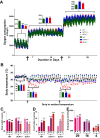
Figure 1.
Figure 1.
Physiological measurements of SLN−/− and UCP1−/− mice during cold adaptation. A, oxygen consumption during housing at 29.0 °C ± 1.0 °C, 16 °C ± 1.0 °C, and 4 °C ± 1.0 °C. The insets show average oxygen consumption for WT, UCP1−/−, and SLN−/− at 29.0 °C ± 1.0 °C, 16 °C ± 1.0 °C, and 4 °C ± 1.0 °C. B, Tc as measured during the entire cold adaptation. Bar graphs show the average Tc during different cold exposure periods. Black arrows under the graphs indicate a switch in ambient temperature. C, body weight in grams at the end of the cold challenge. D, average food consumption per mouse per day in grams. E, ambulatory physical activity (xy axes) measured at different housing temperatures. Each break in the infrared beam is counted as one. One-way ANOVA test for multiple comparisons was performed to analyze statistical difference. The data for the WT at given ambient temperature were treated as a control for statistical analysis. Data from 6 animals/group were analyzed. W, wild type; U, UCP1−/−; S, SLN−/−. No statistical difference is labeled as ns.
Figure 2.
Figure 2.
Cold-induced remodeling of BAT tissue in SLN−/− mice. A, H&E staining of BAT from SLN−/− and WT mouse littermates adapted to 29.0 °C ± 1.0 °C, 16 °C ± 1.0 °C, and 4 °C ± 1.0 °C. B, immunostaining of BAT with UCP1 antibody. C, quantification of UCP1 expression in BAT using the software Halo. The area with positive staining obtained for BAT from the UCP1−/− mice was taken as background and deducted from that obtained for WT and SLN−/− mice. D, representative Western blots of various mitochondrial proteins in BAT. Ponceau S staining is provided as a control to suggest equivalent loading. C1, complex I; C2, complex II; C3, complex III; C4, complex IV; C5, complex V, i.e. ATP synthase. The molecular masses of the detected bands are indicated. E, intensity of few of the proteins that show significant alteration after cold adaptation. Quantification of proteins was performed using ImageJ. W, wild type; U, UCP1−/−; S, SLN−/−. Statistical difference was analyzed using one-way ANOVA for multiple comparisons. No statistical difference is labeled as ns.
Figure 3.
Figure 3.
Electron microscopy of BAT from cold-adapted mice. Representative electron micrographs of BAT from cold-adapted mice are presented. The images presented as top panels and bottom panels for each genotype were obtained at low and high magnification, respectively. Housing temperature is indicated at the top. N, nucleus; L, lipid droplet; M, mitochondria. At thermoneutrality, mitochondria in BAT exhibit a very low abundance of crista structures irrespective of genotype. Interestingly, upon cold exposure, there is an increase in the abundance of mitochondrial cristae in WT and SLN−/− mice (top and bottom rows, respectively) but not in the UCP1−/− littermates.
Figure 4.
Figure 4.
Up-regulation of oxidative metabolism and SLN in the skeletal muscle of UCP1−/− mice during cold adaptation. A, representative images showing SDH staining of skeletal muscle. B, Western blot of SLN showing increased expression after cold adaptation in UCP1−/− mice. Myoglobin and calsequestrin 1 (CASQ1) were used as internal loading controls. C, quantification of SLN expression in oxidative skeletal muscle after adaptation to cold. D, mitochondrial respiration of soleus muscle from WT and SLN−/− mice adapted to severe cold (4 °C). Statistical difference was analyzed using one-way ANOVA for multiple comparisons. No statistical difference is labeled as ns.
Figure 5.
Figure 5.
Increased recruitment of skeletal muscle thermogenesis in UCP1−/− mice. A, transmission electron micrographs of skeletal muscle showing intermyofibrillar mitochondrial abundance in quadriceps muscle. WT and UCP1−/− mice show higher mitochondrial abundance upon adaptation to cold. B, high-magnification electron micrographs showing intermyofibrillar mitochondria with an increased density of crista structures, especially in UCP1−/− mice acclimatized to cold. In contrast, intermyofibrillar mitochondria in muscles from SLN−/− mice do not show any elaboration of crista structures. C, number of mitochondria per low magnification electron micrographs. D, expression of anti-cluster of differentiation 31 (CD31) in skeletal muscle. E, representative image of immunostaining using anti-CD31 (green) and anti-laminin (red) antibodies to probe neovascularization of skeletal muscle. Statistical difference was analyzed using one-way ANOVA for multiple comparisons. No statistical difference is labeled as ns.
Figure 6.
Figure 6.
Increased recruitment of BAT-based thermogenesis in SLN−/− mice during cold adaptation. A, chromogenic staining (brown) of BAT with anti-TH antibody. The location of positive TH staining is shown by black arrows. B, representative images of BAT with anti-CD31 (red) and anti-NPY (brown) antibodies. Positive areas of staining are indicated by black arrows. C, quantification of positively stained areas for TH, NPY, and CD31. Quantification was performed using Halo software. W, wild type; U, UCP1−/−; S, SLN−/−. D, representative Western blotting of NPY and VEGF-R2. Ponceau S staining was provided as a control to indicate equivalent loading for all samples analyzed. E, quantification of NPY and VEGF-R2 protein expression in the BAT. Statistical difference was analyzed using one-way ANOVA for multiple comparisons. No statistical difference is labeled as ns.

References

    1. Oelkrug R., Polymeropoulos E. T., and Jastroch M. (2015) Brown adipose tissue: physiological function and evolutionary significance. J. Comp. Physiol. B 185, 587–606 - PubMed
    1. Cannon B., and Nedergaard J. (2004) Brown adipose tissue: function and physiological significance. Physiol. Rev. 84, 277–359 - PubMed
    1. Azzu V., and Brand M. D. (2010) The on-off switches of the mitochondrial uncoupling proteins. Trends Biochem. Sci. 35, 298–307 - PMC - PubMed
    1. Nedergaard J., Golozoubova V., Matthias A., Asadi A., Jacobsson A., and Cannon B. (2001) UCP1: the only protein able to mediate adaptive non-shivering thermogenesis and metabolic inefficiency. Biochim. Biophys. Acta 1504, 82–106 - PubMed
    1. Fedorenko A., Lishko P. V., and Kirichok Y. (2012) Mechanism of fatty-acid-dependent UCP1 uncoupling in brown fat mitochondria. Cell 151, 400–413 - PMC - PubMed

Publication types

LinkOut - more resources