

White matter hyperintensity reduction and outcomes after minor stroke
- PMID: 28794252
- PMCID: PMC5589793
- DOI: 10.1212/WNL.0000000000004328
White matter hyperintensity reduction and outcomes after minor stroke
Abstract
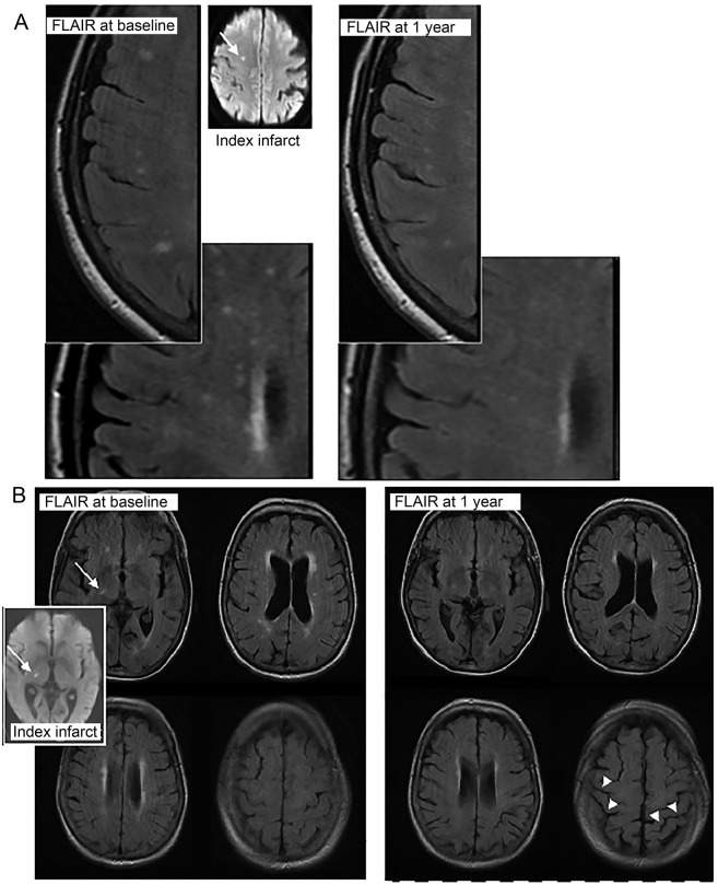
Objective: To assess factors associated with white matter hyperintensity (WMH) change in a large cohort after observing obvious WMH shrinkage 1 year after minor stroke in several participants in a longitudinal study.
Methods: We recruited participants with minor ischemic stroke and performed clinical assessments and brain MRI. At 1 year, we assessed recurrent cerebrovascular events and dependency and repeated the MRI. We assessed change in WMH volume from baseline to 1 year (normalized to percent intracranial volume [ICV]) and associations with baseline variables, clinical outcomes, and imaging parameters using multivariable analysis of covariance, model of changes, and multinomial logistic regression.
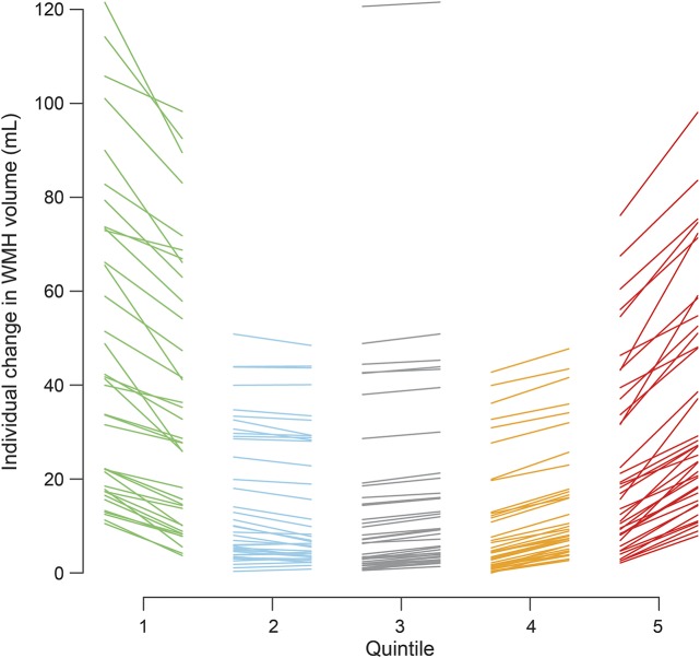
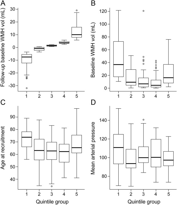
Results: Among 190 participants (mean age 65.3 years, range 34.3-96.9 years, 112 [59%] male), WMH decreased in 71 participants by 1 year. At baseline, participants whose WMH decreased had similar WMH volumes but higher blood pressure (p = 0.0064) compared with participants whose WMH increased. At 1 year, participants with WMH decrease (expressed as percent ICV) had larger reductions in blood pressure (β = 0.0053, 95% confidence interval [CI] 0.00099-0.0097 fewer WMH per 1-mm Hg decrease, p = 0.017) and in mean diffusivity in normal-appearing white matter (β = 0.075, 95% CI 0.0025-0.15 fewer WMH per 1-unit mean diffusivity decrease, p = 0.043) than participants with WMH increase; those with WMH increase experienced more recurrent cerebrovascular events (32%, vs 16% with WMH decrease, β = 0.27, 95% CI 0.047-0.50 more WMH per event, p = 0.018).
Conclusions: Some WMH may regress after minor stroke, with potentially better clinical and brain tissue outcomes. The role of risk factor control requires verification. Interstitial fluid alterations may account for some WMH reversibility, offering potential intervention targets.
Copyright © 2017 The Author(s). Published by Wolters Kluwer Health, Inc. on behalf of the American Academy of Neurology.
Figures
Comment in
-
Waxing and waning of white matter hyperintensities.Neurology. 2017 Sep 5;89(10):984-985. doi: 10.1212/WNL.0000000000004345. Epub 2017 Aug 9. Neurology. 2017. PMID: 28794247 No abstract available.
References
MeSH terms
Grants and funding
LinkOut - more resources
Full Text Sources
Other Literature Sources
Medical