. 2017 Aug 9;7(1):7666.
doi: 10.1038/s41598-017-07977-0.

Complete mitochondrial genomes of Anopheles stephensi and An. dirus and comparative evolutionary mitochondriomics of 50 mosquitoes

Affiliations

Complete mitochondrial genomes of Anopheles stephensi and An. dirus and comparative evolutionary mitochondriomics of 50 mosquitoes

You-Jin Hao et al. Sci Rep. .

Abstract

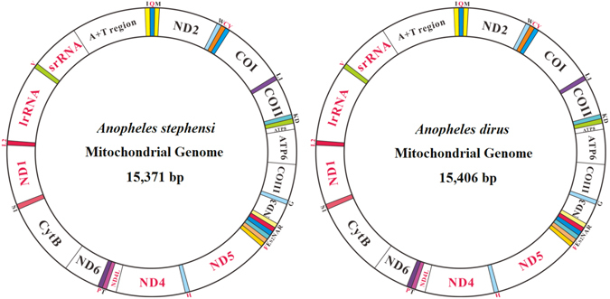
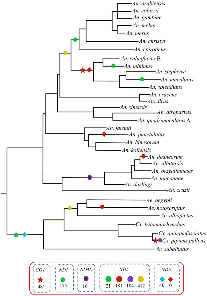
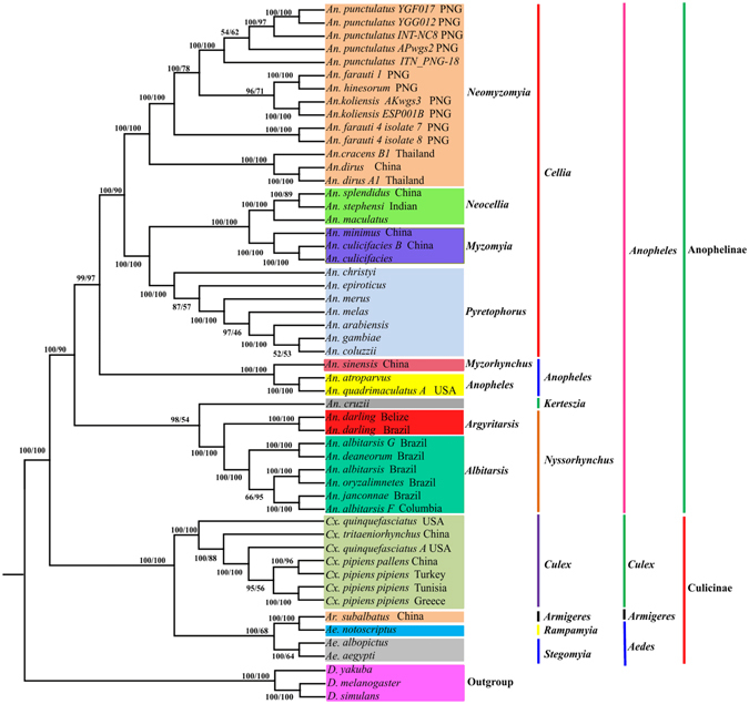
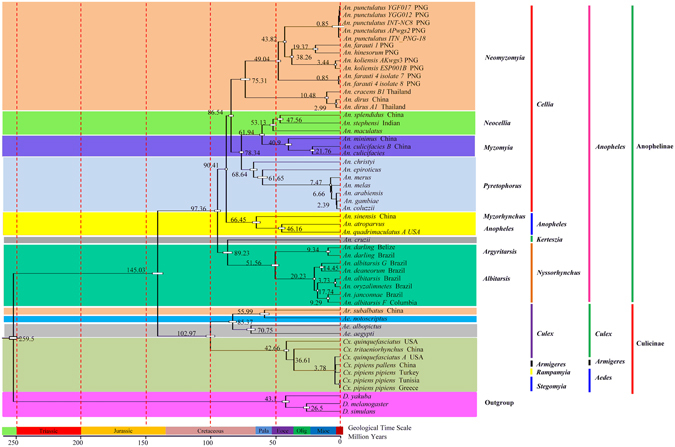
To better understand the phylogeny and evolution of mosquitoes, the complete mitochondrial genome (mitogenome) of Anopheles stephensi and An. dirus were sequenced and annotated, and a total of 50 mosquito mitogenomes were comparatively analyzed. The complete mitogenome of An. stephensi and An. dirus is 1,5371 bp and 1,5406 bp long, respectively. The main features of the 50 mosquito mitogenomes are conservative: 13 protein-coding genes (PCGs), two ribosomal RNA genes, 22 transfer RNA genes, positive AT-skew and negative GC-skew. The gene order trnA-trnR in ancestral insects is rearranged. All tRNA genes have the typical clover leaf secondary structure but tRNA Ser . The control regions are highly variable in size. PCGs show signals of purifying selection, but evidence for positive selection in ND2, ND4 and ND6 is found. Bayesian and Maximum Likelihood phylogenetic analyses based on all PCG nucleotides produce an identical tree topology and strongly support the monophyly of subgenera Cellia, Anopheles, Keterszia and Nyssorhynchus, the sister relationship of the subgenera Nyssorhynchus and Keterszia, and Cellia and Anopheles. The most recent ancestor of the genus Anopheles and Culicini + Aedini exited ~145 Mya ago. This is the first comprehensive study of mosquito mitogenomes, which are effective for mosquito phylogeny at various taxonomic levels.


Conflict of interest statement

The authors declare that they have no competing interests.

Figures

Figure 1
Figure 1
Mitochondrial genome structure of Anopheles stephensi and An. dirus. The color-filled blocks indicate tRNAs, while the un-filled white blocks denote protein-coding genes, rRNA and control regions. The protein-coding genes, rRNA and control regions with black name are located on majority strand, whereas those with red names are on minority strand.
Figure 2
Figure 2
Comparative analysis of AT content, AT- and GC-skew in 50 mosquito mitogenomes. Red, orange, green, light blue, purple, blue and light green dots represent mitogenomes from the subgenus Cellia, Anopheles, Nyssorhynchus, Aedes, Culex, Kertezia and Armigeres, respectively.
Figure 3
Figure 3
Evaluation of codon bias in 50 mosquito mitogenomes. (A) ENC-GC3 plot. The standard curve shows the functional relation between ENC and GC3 under mutation pressure without selection. Points on or close to the curve indicate codon use bias caused by mutation pressure; otherwise, they are affected by natural selection or other factors. (B) Neutrality plot of GC12 versus GC3. GC12 indicates the average value of GC content in the first and second position of the codons (GC1 and GC2); GC3 refers to the GC content in the third position.
Figure 4
Figure 4
The structural organization of the control region of 30 fully completed mosquito mitogenomes. The location and copy number of tandem repeats are shown by colored circle. Non-repeat regions are indicated by colored box with sequence size inside.
Figure 5
Figure 5
Positive selection estimated by FEL site-by-site analyses and pairwise divergence Ka/Ks ratio across mitochondrial protein-coding genes in 50 mosquito mitogenomes. (A) Ratios estimation. Box plot for pairwise divergence of Ka/Ks ratio for each one of the mitochondrial subunits. (B) Percentage of sites under positive selection.
Figure 6
Figure 6
Adaptive evolution analyses based on MEME branch-site selection model. Identification of the maximum likelihood tree branches to test the adaptive evolution of each of the 13 PCGs in 34 representative mosquito species. The putatively positive selection sites with posterior probabilities ≥95% (p-value < 0.05) were marked with different symbols on the represented neighbor-joining tree of ATP6.
Figure 7
Figure 7
Reconstruction of phylogenetic tree determined by RAxML rapid bootstrap and Bayesian inference methods based on all sites (PCG123) of the concatenated protein-coding genes of 50 mosquito mitogenomes. Bayesian posterior probabilities (BPP) (left) and bootstrap support values (BP) are shown at relevant branches of the BI tree.
Figure 8
Figure 8
Evolutionary timescale for subfamily Anophelinae and Culicinae inferred from mtgenome PCG123 datasets based on BI tree. Numbers near the nodes indicate the average divergence time estimated (million years, Mya). In the geological time scale: Pala, Eoce, Olig and Mioc indicate Palaeocene, Eocene, Oligocene and Miocene, respectively.

References

    1. Harbach RE, Kitching IJ. The phylogeny of Anophelinae revisited: inferences about the origin and classification of Anopheles (Diptera: Culicidae) Zoologica Scripta. 2015;45:34–47. doi: 10.1111/zsc.12137. - DOI
    1. Benelli G. Research in mosquito control: current challenges for a brighter future. Parasitology Research. 2015;114:2801–2805. doi: 10.1007/s00436-015-4586-9. - DOI - PubMed
    1. Beebe NW, Cooper RD. Distribution and evolution of the Anopheles punctulatus group (Diptera: Culicidae) in Australia and Papua New Guinea. International Journal for Parasitology. 2002;32:563–574. doi: 10.1016/S0020-7519(01)00359-9. - DOI - PubMed
    1. Krzywinski J, Wilkerson RC, Besansky NJ. Toward understanding Anophelinae (Diptera, Culicidae) phylogeny: Insights from nuclear single-copy genes and the weight of evidence. Systematic Biology. 2001;50:540–556. doi: 10.1080/106351501750435095. - DOI - PubMed
    1. Sallum MAM, et al. Phylogeny of Anophelinae (Diptera: Culicidae) based on nuclear ribosomal and mitochondrial DNA sequences. Systematic Entomology. 2002;27:361–382. doi: 10.1046/j.1365-3113.2002.00182.x. - DOI

Publication types

LinkOut - more resources