. 2017 Aug 9;7(1):7711.
doi: 10.1038/s41598-017-07394-3.

TGFβ signaling is associated with changes in inflammatory gene expression and perineuronal net degradation around inhibitory neurons following various neurological insults

Affiliations

TGFβ signaling is associated with changes in inflammatory gene expression and perineuronal net degradation around inhibitory neurons following various neurological insults

Soo Young Kim et al. Sci Rep. .

Abstract

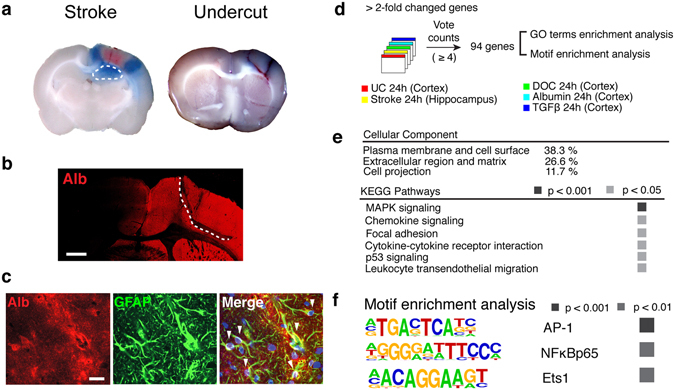
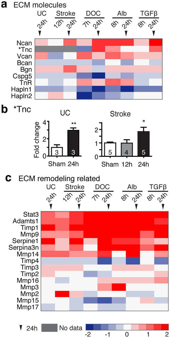
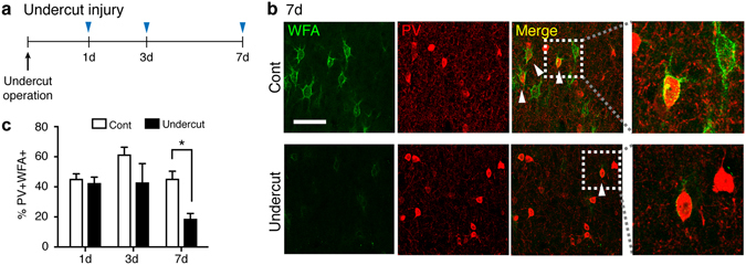
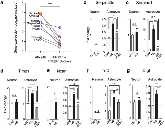
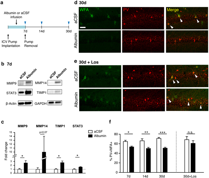
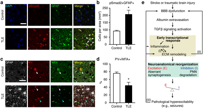
Brain damage due to stroke or traumatic brain injury (TBI), both leading causes of serious long-term disability, often leads to the development of epilepsy. Patients who develop post-injury epilepsy tend to have poor functional outcomes. Emerging evidence highlights a potential role for blood-brain barrier (BBB) dysfunction in the development of post-injury epilepsy. However, common mechanisms underlying the pathological hyperexcitability are largely unknown. Here, we show that comparative transcriptome analyses predict remodeling of extracellular matrix (ECM) as a common response to different types of injuries. ECM-related transcriptional changes were induced by the serum protein albumin via TGFβ signaling in primary astrocytes. In accordance with transcriptional responses, we found persistent degradation of protective ECM structures called perineuronal nets (PNNs) around fast-spiking inhibitory interneurons, in a rat model of TBI as well as in brains of human epileptic patients. Exposure of a naïve brain to albumin was sufficient to induce the transcriptional and translational upregulation of molecules related to ECM remodeling and the persistent breakdown of PNNs around fast-spiking inhibitory interneurons, which was contingent on TGFβ signaling activation. Our findings provide insights on how albumin extravasation that occurs upon BBB dysfunction in various brain injuries can predispose neural circuitry to the development of chronic inhibition deficits.


Conflict of interest statement

The authors declare that they have no competing interests.

Figures

Figure 1
Figure 1
Comparative analysis revealed a common transcriptional signature in hyperexcitable regions following different insults. (a) Evans blue extravasation indicating BBB disruption in the peri-infarct hippocampus (outlined with white dotted line) and around the partially-isolated cortical regions 12 hours after stroke (left) and the undercut operation (right). (b) Representative micrograph of a rat brain slice immunostained for albumin (Alb) at 24 hours following the undercut (UC) operation (indicated by white dotted line). Scale bar = 1mm. (c) Representative confocal images of the UC cortex showing co-localization (white arrowheads) of albumin with GFAP (+) astrocytes. Nuclear staining with DAPI (blue) in the merged image. Scale bar = 20 μm. (d) A vote counting method for five independent microarray datasets from the undercut cortex (UC), peri-infarct hippocampus (Stroke), BBB-disrupted cortex treated with sodium deoxycholate (DOC), and cortices exposed to albumin (Alb) or TGFβ1 (TGFβ). (e) The gene ontology (GO) term enrichment analysis in the common transcriptional profile. (f) Using HOMER software, motifs including AP-1, NFκB, and ETS1 were found significantly enriched in the promoter proximal region, ±500 base pairs of the commonly upregulated genes across models.
Figure 2
Figure 2
Transcriptional activation of TGFβ-regulated extracellular matrix genes across models. The expression levels of genes encoding for extracellular matrix (ECM) components (a) and for molecules involved in ECM remodeling (c) are shown across models. Heat maps are based on a log2 scale. (b) Quantitative real-time PCR was used to measure mRNA level of Tnc in the undercut (UC) cortex and peri-infarct hippocampi (Stroke). One way ANOVA with posthoc Turkey’s test (in Stroke, B) and student t-test (in UC). The number of animals used per condition is indicated within the bar.
Figure 3
Figure 3
Perineuronal nets around PV(+) interneurons are degraded following traumatic brain injury. (a) Time line of experimental design. (b) Representative confocal images of rat undercut and contralateral (Cont) cortices stained for parvalbumin (PV) and perineuronal nets using Wisteria Floribunda agglutinin (WFA) 7 days after the operation. Scale bar = 50 µm. White arrowheads indicate representative PV(+)/WFA(+) cells. Areas within white rectangles in merged images are shown magnified. (c) The percentage of PV(+) interneurons associated with PNNs was significantly decreased at 7 days following the undercut operation. *p < 0.05, Two-way ANOVA with post-hoc Sidak’s test, two tailed. Three biological replicates (n’s = 3) were used at each time point.
Figure 4
Figure 4
Albumin activated the transcriptional responses of ECM genes via TGFβ receptor signaling. (a) The expression levels of ECM-related genes is upregulated across models (marked in bold in A, C) in a separate microarray data set in comparisons between albumin vs. albumin + TGFβ receptor (R) blockers. **p = 0.0078, Wilcoxon signed-rank test, two-tailed. (bg) Quantitative real-time PCR for the mRNA expression of selected genes in primary cortical astrocytic or neuronal cultures at 24 hours following treatment of albumin or albumin plus a specific Alk5 blocker, SJN2511. Results were of three independent primary culture derivations (n = 11 per condition in neurons; n = 10–12 per condition in astrocytes). One way ANOVA with posthoc Turkey’s test (in astrocytes) and student t-test (in neurons) were performed. Cont, control; Alb, albumin; SJN, SJN2511; n.d., non detectable (Cq value ≥ 35). *p < 0.05, **p < 0.01, ***p < 0.001, ****p < 0.0001. Data are shown as mean ± S.E.
Figure 5
Figure 5
Albumin induced the degradation of perineuronal nets around PV(+) interneurons and losartan attenuated the effect of albumin. (a) Experimental design. Intracerebroventricular (ICV) osmotic pumps containing albumin (0.4 mM) or aCSF were implanted in rats and mice and removed after 7 days. (b,c) Molecules involved in ECM remodeling including MMP9, TIMP1, and STAT3 were significantly increased in dissected rat hippocampi following albumin infusion for 7 days. Uncropped blot images are available in the Supplementary Fig. S4. Each group has four biological replicates. Unpaired student t- test. *p < 0.05, two-tailed. (d) Representative confocal images showing PNNs (WFA) and PV(+) cells in the mouse hippocampal CA1 region at 30 days post implantation. (e) A separate set of animals was infused with albumin + losartan (10μM) or aCSF + losartan. Scale bar = 50 μm. (f) Albumin exposure decreased the association of PNNs with PV(+) interneurons (*p < 0.05, **p < 0.01, ***p < 0.001, Two-way ANOVA, post-hoc Sidak’s test, two tailed). Co-administration of losartan attenuated the effect of albumin (p > 0.05, Mann-Whitney test, two-tailed). n.s., not significant. Each condition has three or four biological replicates (n = 3–4).
Figure 6
Figure 6
Increased expression of astrocytic pSmad2 and reduced association of PNNs around PV(+) cells in human epileptic hippocampi. (a) Representative micrographs of human hippocampi stained for GFAP and phosphorylated Smad2, a downstream effecter of TGFβ signaling. The hippocampi were resected from temporal lobe epilepsy (TLE) patients or age-matched autopsy controls. Representative GFAP(+) astrocytes expressing pSmad2(+) are indicated by white arrowheads. Scale bar = 50 μm. (b) Co-localization of pSmad2 with GFAP was increased in TLE patients (n = 5) compared to controls (n = 3). (c) Representative confocal micrographs of human hippocampal tissues stained for GFAP, PV, and PNNs (WFA) resected from TLE patients or controls. Scale bar = 50 μm. White and blue arrowheads indicate representative PV(+)/WFA(+) and PV(+)/WFA(−) cells, respectively. (d) The percentage of PV(+)/WFA(+) cells was decreased in the hippocampus of TLE patients (n = 4) compared to controls (n = 4). Mann-Whitney test, two-tailed, was used for statistical analyses. *p < 0.05. (e) A working model. Several points for feed-forward loops (i-iii) can lead to chronic hyperexcitability and the development of epilepsy. The dysfunction of blood-brain barrier (BBB) and the ensuing entry of albumin into brain parenchyma activate TGFβ signaling. Comparative transcriptome analyses predicted the activation of the common core signaling transduction including MAPK pathway, Stat3, NFκB, AP-1, and ETS1. These signaling pathways can elicit the reciprocal activation of inflammation and extracellular matrix (ECM) remodeling (i). ECM remodeling can trigger the transformation of a latent from of TGFβ to its active form as well as exacerbate BBB dysfunction (ii). The degradation of perineuronal nets (PNNs) around fast-spiking interneurons and aberrant excitatory synaptogenesis occur in the course of ECM remodeling, presumably leading to functional alterations in inhibition and abnormality in synaptic plasticity that may contribute to excitation/inhibition (E/I) imbalance and ultimately the occurrence of seizures. Finally, seizures per se cause BBB dysfunction, inflammation, and the upregulation of MMPs activity, (iii).

References

    1. Balami JS, Chen RL, Grunwald IQ, Buchan AM. Neurological complications of acute ischaemic stroke. Lancet Neurol. 2011;10:357–371. doi: 10.1016/S1474-4422(10)70313-6. - DOI - PubMed
    1. Shlosberg D, Benifla M, Kaufer D, Friedman A. Blood-brain barrier breakdown as a therapeutic target in traumatic brain injury. Nature Reviews Neurology. 2010;6:393–403. doi: 10.1038/nrneurol.2010.74. - DOI - PMC - PubMed
    1. Abbott NJ. Astrocyte-endothelial interactions and blood-brain barrier permeability. J Anat. 2002;200:629–638. doi: 10.1046/j.1469-7580.2002.00064.x. - DOI - PMC - PubMed
    1. Bar-Klein G, et al. Losartan prevents acquired epilepsy via TGF-beta signaling suppression. Ann Neurol. 2014;75:864–875. doi: 10.1002/ana.24147. - DOI - PMC - PubMed
    1. Seiffert E, et al. Lasting blood-brain barrier disruption induces epileptic focus in the rat somatosensory cortex. J Neurosci. 2004;24:7829–7836. doi: 10.1523/JNEUROSCI.1751-04.2004. - DOI - PMC - PubMed

Publication types

MeSH terms

Substances