

Mycetoma: An Update
- PMID: 28794542
- PMCID: PMC5527712
- DOI: 10.4103/ijd.IJD_476_16
Mycetoma: An Update
Abstract
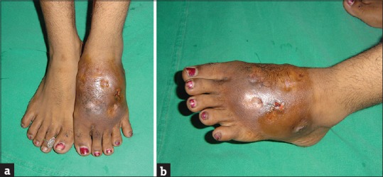
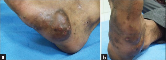
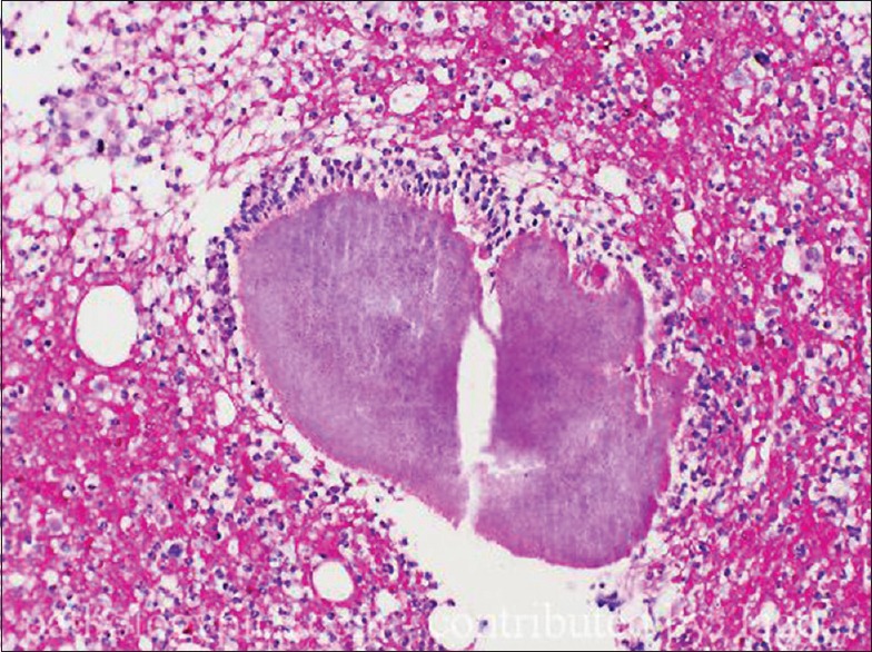
Mycetoma is a localized chronic, suppurative, and deforming granulomatous infection seen in tropical and subtropical areas. It is a disorder of subcutaneous tissue, skin and bones, mainly of feet, characterized by a triad of localized swelling, underlying sinus tracts, and production of grains or granules. Etiological classification divides it into eumycetoma caused by fungus, and actinomycetoma caused by bacteria. Since the treatment of these two etiologies is entirely different, a definite diagnosis after histopathological and microbiological examination is mandatory, though difficult. Serological test exists but is not so reliable; however, molecular techniques to identify relevant antigens have shown promise. The disease is notoriously difficult to treat. Eumycetoma may be unresponsive to standard antifungal therapy. Actinomycetoma responds to antibiotic therapy, but prolonged treatment is necessary. This review focuses on the etiopathogenesis, clinical features, laboratory diagnosis, and treatment of mycetoma.
Keywords: Actinomycetoma; Madura foot; eumycetoma.
Conflict of interest statement
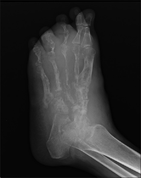
There are no conflicts of interest. What is new? Greater frequency of disease in men may not be just attributable to environmental factors but also to hormonal factorsVarious serological and molecular tests have also found place in diagnosis of mycetoma allowing early diagnosis and identification of new species and phylogenetic relationshipsMagnetic resonance imaging provides the most comprehensive method for assessment of the bone and soft tissue involvement and may also be useful in evaluating the differential diagnosis of the swellingCombination antibiotic therapy is a must in case of actinomycetomasSeveral newer antifungals have been tried for eumycetomas though in vivo studies are lacking.
Figures
References
-
- McGinnis MR. Mycetoma. Dermatol Clin. 1996;14:97–104. - PubMed
-
- Kwon-Chung KJ, Bennett JE. Medical Mycology. Philadelphia: Lea & Febiger; 1992. Mycetoma; pp. 560–93.
-
- Carter HV. On a New and Striking form of Fungus Disease Principally Affecting the Foot and Prevailing Endemically in Many Parts of India. Transactions of the Medical and Physical Society of Bombay. 1860;6:104–42.
-
- Pinoy E. Actinomycoses and mycetomas. Bull Inst Pasteur. 1913;11:929.
Publication types
LinkOut - more resources
Full Text Sources
Other Literature Sources