. 2017 Jul 26:11:2179-2191.
doi: 10.2147/DDDT.S137507. eCollection 2017.

Schisandrin B attenuates CCl4-induced liver fibrosis in rats by regulation of Nrf2-ARE and TGF-β/Smad signaling pathways

Affiliations

Schisandrin B attenuates CCl4-induced liver fibrosis in rats by regulation of Nrf2-ARE and TGF-β/Smad signaling pathways

Qingshan Chen et al. Drug Des Devel Ther. .

Abstract

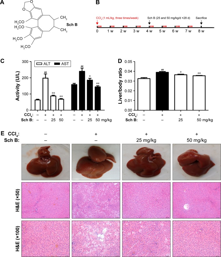
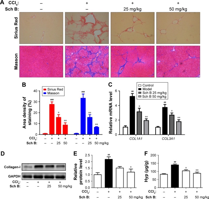
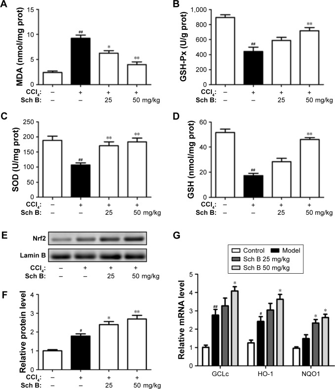
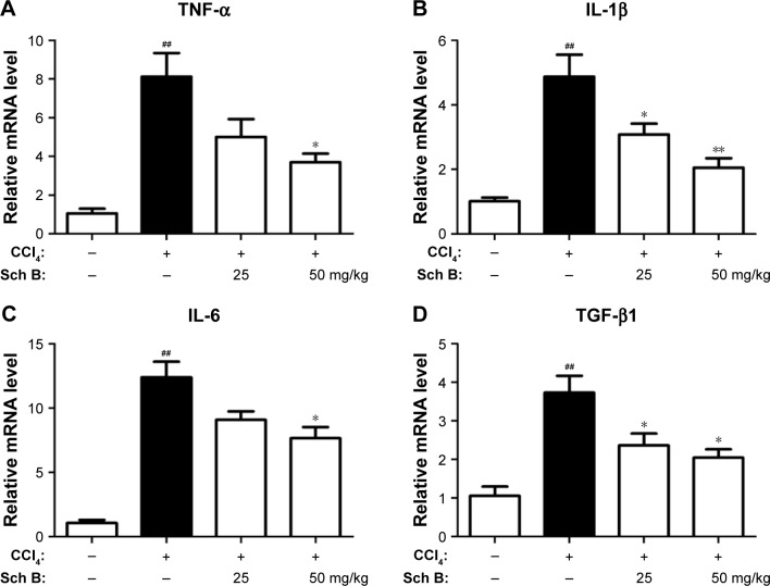
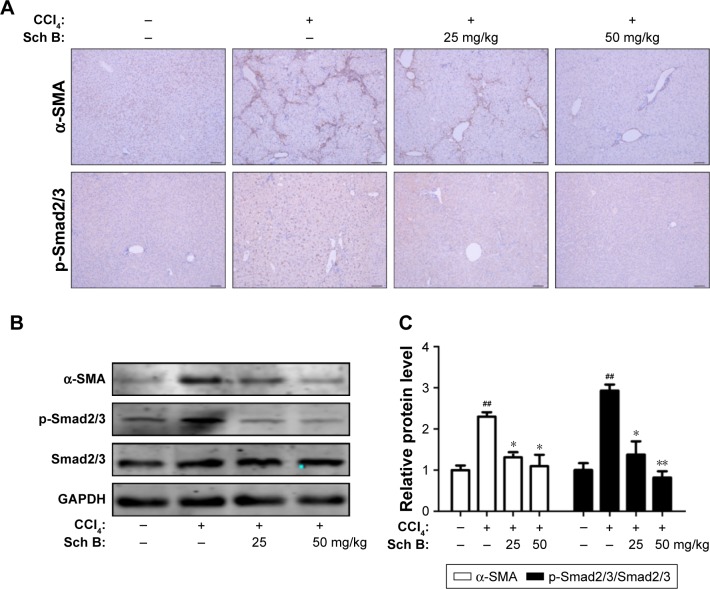
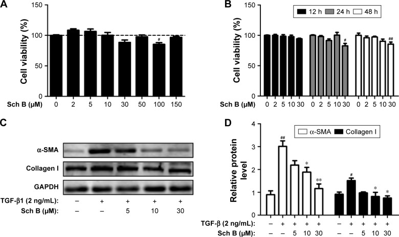
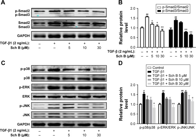
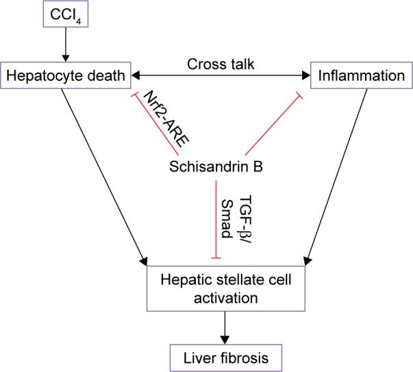
Liver fibrosis is a major pathological feature of chronic liver diseases and there is no effective therapy program at present. Schisandrin B (Sch B) is the major bioactive ingredient of Schisandra chinensis, with antioxidative, anti-inflammatory, antitumor, and hepatoprotective properties. This study aimed to investigate the protective effect and related molecular mechanism of Sch B against carbon tetrachloride (CCl4)-induced liver fibrosis in rats. The in vivo therapeutic effect of Sch B on liver fibrosis induced by CCl4 was examined in rats. In vitro, rat hepatic stellate cells (HSC-T6) were used to assess the effect of Sch B on the activation of HSCs. Sch B effectively attenuated liver damage and progression of liver fibrosis in rats, as evidenced by improved liver function and decreased collagen deposition. The effects of Sch B were associated with attenuating oxidative stress by activating nuclear factor-erythroid 2-related factor 2 (Nrf2)-mediated antioxidant signaling and suppressing HSC activation by inhibiting the transforming growth factor-β (TGF-β)/Smad signaling pathway. In an in vitro study, it was shown that Sch B inhibited TGF-β-induced HSC activation. Finally, Sch B significantly inhibited TGF-β1-stimulated phosphorylation of Smad and signaling of mitogen-activated protein kinases. This study demonstrates that Sch B prevents the progression of liver fibrosis by the regulation of Nrf2-ARE and TGF-β/Smad signaling pathways, and indicates that Sch B can be used for the treatment of liver fibrosis.

Keywords: Nrf2; TGF-β/Smad; hepatic stellate cell activation; liver fibrosis; schisandrin B.


Conflict of interest statement

Disclosure The authors report no conflicts of interest in this work.

Figures

Figure 1
Figure 1
Sch B protects against CCl4-induced liver injury. Notes: (A) The structure of Schisandrin B (Sch B). (B) The experimental design scheme. (C) Serum ALT and AST activities. (D) Liver weight to body weight ratio. (E) H&E staining of liver sections (Scale bar: 100 and 50 μm; original magnification, ×50 and ×100). Data are expressed as the mean ± SEM (n=8). ##P<0.01 versus control group. *P<0.05 and **P<0.01 versus model group. Abbreviations: CCl4, carbon tetrachloride; d, days; w, weeks; ALT, alanine transaminase; AST, aspartate transaminase.
Figure 2
Figure 2
Sch B ameliorates CCl4-induced liver fibrosis in rats. Notes: (A, B) Liver fibrosis assessed by Sirius Red and Masson. (C) The mRNA levels of COL1A1 and COL3A1. (D) Western blot analysis of collagen-I. (E) Densitometric analysis of Western blots. (F) Hydroxyproline content in rat livers. Data are expressed as the mean ± SEM (n=8). ###P<0.001 and ##P<0.01 versus control group. *P<0.05 and **P<0.01 versus model group. Abbreviations: CCl4, carbon tetrachloride; Sch B, schisandrin B; Hyp, hydroxyproline.
Figure 3
Figure 3
Sch B regulates CCl4-induced hepatic oxidative stress and Nrf2-ARE signaling pathway in vivo. Notes: (A) Lipid peroxidation MDA. (B) Liver GSH-Px activities. (C) Antioxidant enzyme SOD. (D) Liver GSH levels. (E) Western blot analysis of Nrf2 levels in the liver. (F) Densitometric analysis of Western blots. (G) The mRNA levels of GCLc, HO-1, and NQO1. Data are expressed as the mean ± SEM. #P<0.05 and ##P<0.01 versus control group. *P<0.05 and **P<0.01 versus model group. Abbreviations: ARE, antioxidant response element; CCl4, carbon tetrachloride; Nrf2, nuclear factor-erythroid 2-related factor 2; Sch B, schisandrin B; MDA, malondialdehyde; SOD, superoxide dismutase; GSH, glutathione; GSH-Px, glutathione peroxidase.
Figure 4
Figure 4
Sch B alleviates CCl4-induced inflammatory response. Notes: (A) Relative mRNA levels of TNF-α. (B) Relative mRNA levels of IL-1β. (C) Relative mRNA levels of IL-6. (D) Relative mRNA levels of TGF-β1. Data are expressed as the mean ± SEM (n=6). ##P<0.01 versus control group. *P<0.05 and **P<0.01 versus model group. Abbreviations: CCl4, carbon tetrachloride; IL-6, interleukin-6; TGF-β, transforming growth factor-β; TNF-α, tumor necrosis factor-α; Sch B, schisandrin B.
Figure 5
Figure 5
Sch B suppresses HSC activation and TGF-β/Smad signaling in CCl4-induced liver fibrosis. Notes: (A) Immunohistochemistry staining of α-SMA and p-Smad2/3 in the liver tissues, the sections were photographed at Scale bar: 100 μm; original magnification: ×50; (B) Effects of Sch B on the expression of α-SMA, p-Smad2/3, and Smad2/3 in the liver tissues were measured by Western blot analysis; (C) Densitometric analysis of Western Blots. Data are expressed as the mean ± SEM. ##P<0.01 versus control group. *P<0.05 and **P<0.01 versus model group. Abbreviations: CCl4, carbon tetrachloride; HSC, hepatic stellate cell; TGF-β, transforming growth factor β; Sch B, schisandrin B.
Figure 6
Figure 6
Sch B inhibits the TGF-β1-induced HSC activation in vitro. Notes: (A, B) Effects of Sch B on HSC-T6 cells viability. Cell viability was assessed by CCK8 assay; (C) Effects of Sch B on TGF-β1-induced HSC activation as assessed by α-SMA and collagen I expression; (D) Densitometric analysis of Western Blots. Data are expressed as the mean ± SEM. #P<0.05 and ##P<0.01 versus control group. *P<0.05 and **P<0.01 versus model group. Abbreviations: α-SMA, α-smooth muscle actin; HSC, hepatic stellate cell; TGF-β, transforming growth factor-β; Sch B, schisandrin B.
Figure 7
Figure 7
Sch B inhibits TGF-β-mediated Smad and MAPK signaling molecules in vitro. Notes: (A, B) The expression of p-Smad2 and p-Smad3 was assessed by Western blot, and the densitometric analysis of Western blots. (C, D) Western blot analysis of p-p38, p38, p-ERK, ERK, p-JNK, and JNK in HSCs, and the densitometric analysis of Western blots. Data are expressed as the mean ± SEM. #P<0.05 and ##P<0.01 versus control group. *P<0.05 and **P<0.01 versus model group. Abbreviations: HSCs, hepatic stellate cells; MAPK, mitogen-activated protein kinases; TGF-β, transforming growth factor-β; Sch B, schisandrin B.
Figure 8
Figure 8
A diagram shows the possible mechanisms of Sch B against CCl4-induced liver fibrosis in rats. Abbreviations: ARE, antioxidant response element; CCl4, carbon tetrachloride; Nrf2, nuclear factor-erythroid 2-related factor 2; Sch B, schisandrin B.

Similar articles

Cited by

References

    1. Bataller R, Brenner DA. Liver fibrosis. J Clin Invest. 2005;115(2):209–218. - PMC - PubMed
    1. Hernandez-Gea V, Friedman SL. Pathogenesis of liver fibrosis. Ann Rev Pathol. 2011;6:425–456. - PubMed
    1. Zhang DY, Friedman SL. Fibrosis-dependent mechanisms of hepatocarcinogenesis. Hepatology. 2012;56(2):769–775. - PMC - PubMed
    1. Schuppan D, Kim YO. Evolving therapies for liver fibrosis. J Clin Invest. 2013;123(5):1887–1901. - PMC - PubMed
    1. Iwaisako K, Jiang C, Zhang M, et al. Origin of myofibroblasts in the fibrotic liver in mice. Proc Natl Acad Sci U S A. 2014;111(32):E3297–E3305. - PMC - PubMed

MeSH terms