. 2017 Jul 20;5(7):2325967117714998.
doi: 10.1177/2325967117714998. eCollection 2017 Jul.

Distal Musculotendinous T Junction Injuries of the Biceps Femoris: An MRI Case Review

Affiliations

Distal Musculotendinous T Junction Injuries of the Biceps Femoris: An MRI Case Review

Tom Entwisle et al. Orthop J Sports Med. .

Abstract

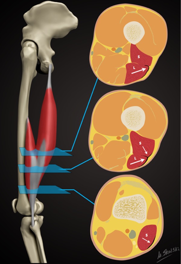
Background: Injury to the distal musculotendinous T junction (DMTJ) of the biceps femoris is a distinct clinical entity that behaves differently from other hamstring injuries due to its complex, multicomponent anatomy and dual innervation. Injury in this region demonstrates a particularly high rate of recurrence, even with prolonged rehabilitation times.

Purpose: To describe the anatomy of the DMTJ of the biceps femoris and analyze the injury patterns seen on magnetic resonance imaging (MRI) to aid prognosis and rehabilitation and minimize the risk of recurrence.

Study design: Cross-sectional study; Level of evidence, 3.

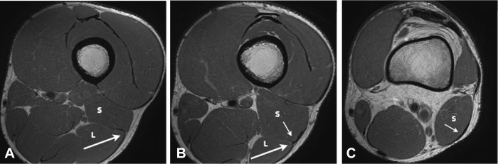
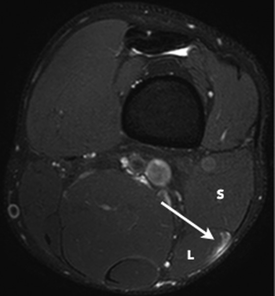
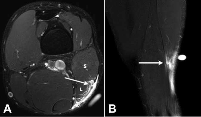
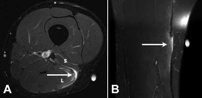
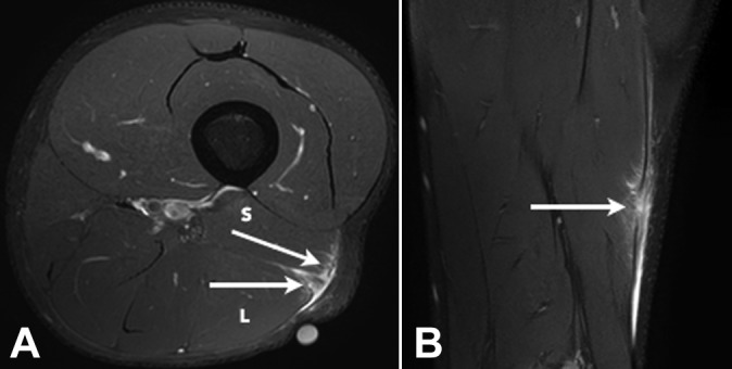
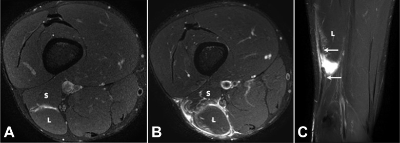
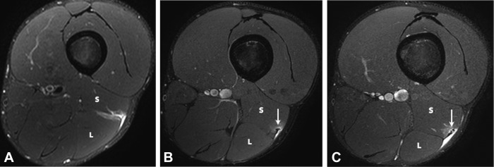
Methods: Acute injury to the DMTJ of the biceps femoris was identified in 106 MRI examinations from 55 patients at a single institution. Each injury was classified as involving the long head, the short head, or both components of the DMTJ, with each component individually graded. Injuries were classified as recurrent if there was a previous MRI demonstrating an acute injury to the DMTJ or if there was scarring present at the site of an acute injury.

Results: Of the 106 acute injuries to the DMTJ of the biceps femoris, isolated injury to the long head component was the most common (51%), with both components involved in [round 42.5% to 43%] of cases. Isolated injury to the short head component accounted for 7% of cases. The recurrence rate for reinjury to the DMTJ was 54% in this series. The date of prior injury was known in 45 of 57 recurrent cases, with 34 of these reoccurring within 3 months (76%) and 40 reoccurring within 12 months (89%). The recurrent injury was of a higher grade than the prior injury in 22 of 44 instances (50%), the same grade in 16 instances (36%), and a lower grade in 6 instances (14%). Thus, 86% of recurrent injuries were of the same or higher grade than prior injury.

Conclusion: These results suggest that high-risk muscle injuries, such as that to the DMTJ of the biceps femoris, should be evaluated using MRI to determine the structural components involved and to assess the extent and severity of injury.

Keywords: biceps femoris; hamstring; magnetic resonance imaging; muscle injury.


Conflict of interest statement

The authors declared that they have no conflicts of interest in the authorship and publication of this contribution.

Figures

Figure 1.
Figure 1.
Schematic demonstrating the sequential axial anatomy of the distal musculotendinous T junction (DMTJ) of the biceps femoris. Proximally, the anterolateral epimysial surface of the long head (L) condenses to form the proximal portion of the DMTJ and appears L-, C-, or U-shaped (large arrow). In the midportion, the opposing epimysial condensations at the anterolateral aspect of the long head and the posterolateral aspect of the short head (S, small arrow) form the DMTJ that appears as a T-shaped structure. Distally, the posterolateral epimysial condensation of the short head forms the DMTJ and appears as a shallow convex structure.
Figure 2.
Figure 2.
Axial proton density images demonstrating the anatomy of the distal musculotendinous T junction (DMTJ) of the biceps femoris. (A) The anterolateral epimysial surface of the long head (L) condenses to form the proximal portion of the DMTJ and appears L-, C-, or U-shaped (large arrow). (B) The opposing epimysial condensations at the anterolateral aspect of the long head (L, large arrow) and the posterolateral aspect of the short head (S, small arrow) form the midportion of the DMTJ that appears T-shaped. (C) The posterolateral epimysial condensation of the short head (S, small arrow) forms the distal portion of the DMTJ and appears as a shallow convex structure.
Figure 3.
Figure 3.
Axial T2 SPAIR (spectral adiabatic inversion recovery) image of a grade 1 strain of the long head component (L) of the distal musculotendinous T junction of the biceps femoris. Note the intact T junction tendon structure with peritendinous edema (arrow) at the myotendinous interface of the long head. The short head component (S) is normal.
Figure 4.
Figure 4.
(A) Axial and (B) coronal T2 SPAIR (spectral adiabatic inversion recovery) images of a grade 2 tear of the long head component of the distal musculotendinous T junction of the biceps femoris. There is a partial-thickness tear of the long head component (L) of the T junction tendon structure (arrows) and peritendinous edema and blood fluid.
Figure 5.
Figure 5.
(A) Axial and (B) coronal T2 SPAIR (spectral adiabatic inversion recovery) images of a grade 3 tear of the long head component (L) of the distal musculotendinous T junction of the biceps femoris. There is a full-thickness tear with retraction of the long head component of the T junction tendon structure (arrows) and peritendinous edema and blood fluid present. The short head component (S) is normal.
Figure 6.
Figure 6.
(A) Axial and (B) coronal T2 SPAIR (spectral adiabatic inversion recovery) image of a grade 2 tear of both the long (L) and short (S) head components of the distal musculotendinous T junction of the biceps femoris with several partial-thickness defects in both the long and short head components of the T junction tendon structure (arrows). Peritendinous edema and blood fluid are noted.
Figure 7.
Figure 7.
(A, B) Axial and (C) coronal T2-weighted T2 SPAIR (spectral adiabatic inversion recovery) images of a complex multicomponent tear in the distal aspect of the biceps femoris muscle. There is a grade 3 full-thickness tear with retraction of the long head component of the distal musculotendinous T junction (DMTJ) (large arrows). Partial-thickness tears of the short head component of the DMTJ (S) and the distal-most aspect of the proximal intramuscular tendon of the long head (small arrows) are also present.
Figure 8.
Figure 8.
(A) Axial T2 SPAIR (spectral adiabatic inversion recovery) image of an initial grade 1 strain of the long (L) and short (S) head components of the distal musculotendinous T junction (DMTJ) of the biceps femoris muscle. (B) Axial and (C) coronal T2 SPAIR images of a subsequent grade 3 tear of the long (L) and short (S) head components of the DMTJ of the biceps femoris muscle in the same patient as in (A). Blood fluid fills the interval between the retracted ends of the tendon (arrows).
Figure 9.
Figure 9.
Axial T2 SPAIR (spectral adiabatic inversion recovery) images of 3 separate injuries involving the distal musculotendinous T junction of the biceps femoris over a 5-week period. The initial injury (A) was a grade 1 strain of both the long (L) and short (S) head components. There is a small amount of muscle fiber disruption at the myotendinous interface of the short head component but the T junction tendon structure was intact throughout. The first recurrent injury (B) was an isolated grade 1 strain of the short head component. A small focus of developing scarring is present at the epicenter of the strain (arrow). The long head component (L) is normal. The second recurrent injury (C) was a grade 2 tear of the short head component, where there is a small longitudinal split/delamination evident (arrow). The long head component (L) is normal.
Figure 10.
Figure 10.
Axial T2 SPAIR (spectral adiabatic inversion recovery) images of 2 separate injuries involving the distal musculotendinous T junction of the biceps femoris over a 4-week period. The initial injury (A) was a grade 1 strain of the long head component (L) and the adjacent posterior epimysium (small arrow). The T junction tendon structure was intact. The recurrent injury (B) demonstrates a full-thickness grade 3 tear with retraction of the long head component and a grade 2 partial-thickness tear of the short head component (large arrows).

References

    1. Ahmad CS, Redler LH, Ciccotti MG, Maffulli N, Longo UG, Bradley J. Evaluation and management of hamstring injuries. Am J Sports Med. 2013;41:2933–2947. - PubMed
    1. Arnason A, Sigurdsson SB, Gudmundsson A, Holme I, Engebretsen L, Bahr R. Risk factors for injuries in football. Am J Sports Med. 2004;32(suppl 1):5S–16S. - PubMed
    1. Branch EA, Anz AW. Distal insertions of the biceps femoris: a quantitative analysis. Orthop J Sports Med. 2015;3:2325967115602255. - PMC - PubMed
    1. Brooks JH, Fuller CW, Kemp SP, Reddin DB. Incidence, risk, and prevention of hamstring muscle injuries in professional rugby union. Am J Sports Med. 2006;34:1297–1306. - PubMed
    1. Brukner P. Hamstring injuries: prevention and treatment—an update. Br J Sports Med. 2015;49:1241–1244. - PMC - PubMed

LinkOut - more resources