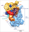
A new era of studying p53-mediated transcription activation
- PMID: 28795863
- PMCID: PMC5834226
- DOI: 10.1080/21541264.2017.1345354
A new era of studying p53-mediated transcription activation
Abstract
To prevent tumorigenesis, p53 stimulates transcription by facilitating the recruitment of the transcription machinery on target gene promoters. Cryo-Electron Microscopy studies on p53-bound RNA Polymerase II (Pol II) reveal that p53 structurally regulates Pol II to affect its DNA binding and elongation, providing new insights into p53-mediated transcriptional regulation.
Keywords: 3D structure; RNA polymerase II; cryo-electron microscopy; p53; transcription activation.
Figures


References
-
- Menendez D, Inga A, Resnick MA. The expanding universe of p53 targets. Nat Rev Cancer 2009; 9:724-37; PMID:19776742; http://doi.org/10.1038/nrc2730 - DOI - PubMed
-
- Bieging KT, Mello SS, Attardi LD. Unravelling mechanisms of p53-mediated tumour suppression. Nat Rev Cancer 2014; 14:359-70; PMID:24739573; http://doi.org/10.1038/nrc3711 - DOI - PMC - PubMed
-
- Kamada R, Toguchi Y, Nomura T, Imagawa T, Sakaguchi K. Tetramer formation of tumor suppressor protein p53: Structure, function, and applications. Biopolymers 2016; 106:598-612; PMID:26572807; http://doi.org/10.1002/bip.22772 - DOI - PubMed
-
- Levine M, Cattoglio C, Tjian R. Looping back to leap forward: transcription enters a new era. Cell 2014; 157:13-25; PMID:24679523; http://doi.org/10.1016/j.cell.2014.02.009 - DOI - PMC - PubMed
-
- Roeder RG. The role of general initiation factors in transcription by RNA polymerase II. Trends Biochem Sci 1996; 21:327-35; PMID:8870495; http://doi.org/10.1016/S0968-0004(96)10050-5 - DOI - PubMed
Publication types
MeSH terms
Substances
Grants and funding
LinkOut - more resources
Full Text Sources
Other Literature Sources
Research Materials
Miscellaneous
