. 2017 Aug 10;9(8):858.
doi: 10.3390/nu9080858.

Suppression of Wnt Signaling and Osteogenic Changes in Vascular Smooth Muscle Cells by Eicosapentaenoic Acid

Affiliations

Suppression of Wnt Signaling and Osteogenic Changes in Vascular Smooth Muscle Cells by Eicosapentaenoic Acid

Yukihiro Saito et al. Nutrients. .

Abstract

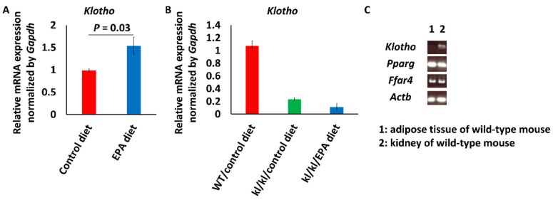
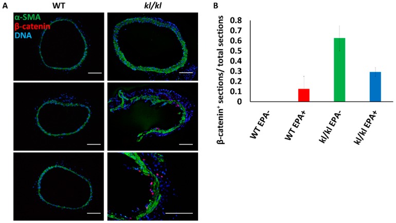
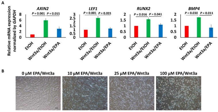
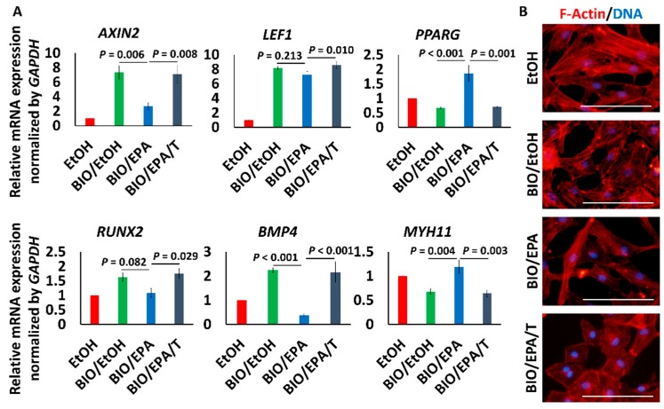
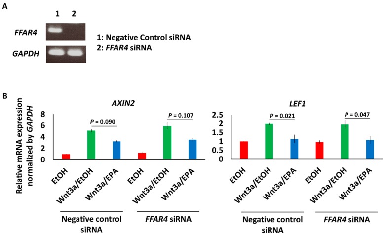
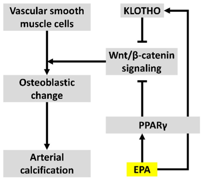
Vascular medial calcification is often observed in patients with arteriosclerosis. It is also associated with systolic hypertension, wide pulse pressure, and fluctuation of blood pressure, which results in cardiovascular events. Eicosapentaenoic acid (EPA) has been shown to suppress vascular calcification in previous animal experiments. We investigated the inhibitory effects of EPA on Wnt signaling, which is one of the important signaling pathways involved in vascular calcification. Intake of food containing 5% EPA resulted in upregulation of the mRNA expression of Klotho, an intrinsic inhibitor of Wnt signaling, in the kidneys of wild-type mice. Expression levels of β-catenin, an intracellular signal transducer in the Wnt signaling pathway, were increased in the aortas of Klotho mutant (kl/kl) mice compared to the levels in the aortas of wild-type mice. Wnt3a or BIO, a GSK-3 inhibitor that activates β-catenin signaling, upregulated mRNA levels of AXIN2 and LEF1, Wnt signaling marker genes, and RUNX2 and BMP4, early osteogenic genes, in human aorta smooth muscle cells. EPA suppressed the upregulation of AXIN2 and BMP4. The effect of EPA was cancelled by T0070907, a PPARγ inhibitor. The results suggested that EPA could suppress vascular calcification via the inhibition of Wnt signaling in osteogenic vascular smooth muscle cells via PPARγ activation.

Keywords: Wnt signaling; eicosapentaenoic acid; vascular calcification.


Conflict of interest statement

The authors have no conflict of interest to declare.

Figures

Figure 1
Figure 1
Klotho (kl/kl) mRNA expression levels in kidneys of mice. (A) Klotho mRNA expression levels in kidneys of wild-type mice fed a diet with or without eicosapentaenoic acid (EPA). Data are shown as means ± SE (Control diet, n = 4; EPA diet, n = 8) and were analyzed using Student’s t test; (B) Klotho mRNA expression levels in kidneys of kl/kl mice fed a diet with or without EPA. Data are shown as means (n = 2 in each group); (C) Reverse transcription-polymerase chain reaction (RT-PCR) showed the mRNA of EPA receptors, Pparg and Ffar4, in the kidney.
Figure 2
Figure 2
Beta-catenin expression in aortas of mice. (A) Immunostaining of mouse aortas: green, α-smooth muscle actin (α-SMA); red, β-catenin; and blue, DNA; (B) Proportions of β-catenin-positive aorta sections (total numbers of sections: Wild-type (WT) mice fed control diet, n = 7; WT mice fed EPA diet, n = 6; kl/kl mice fed control diet, n = 6; and kl/kl mice fed EPA diet, n = 7). Bar = 100 μm.
Figure 3
Figure 3
Effects of Wnt3a and EPA on human aortic smooth muscle cells (HAoSMCs). (A) Changes of the gene expression pattern in HAoSMCs treated with or not treated with 100 ng/mL Wnt3a and/or EPA. Data are shown as means ± SE (n = 4 in each group), and were analyzed using one-way ANOVA with Tukey’s post hoc test. EtOH means ethanol; (B) Cytotoxicity of EPA in HAoSMCs. Bar = 100 μm.
Figure 4
Figure 4
Effects of EPA on β-catenin signaling stimulated by GSK-3 inhibitor. (A) Changes of the gene expression pattern in HAoSMCs treated with 1 μmol/L BIO, with or without EPA/T0070907. Data are shown as means ± SE (n = 4 in each group) and were analyzed using one-way ANOVA with Tukey’s post hoc test. T means T0070907 (10 μmol/L); (B) Morphological changes of HAoSMCs. Red and blue represent f-actin and DNA, respectively. Bar = 100 μm.
Figure 5
Figure 5
Effect of the knockdown of FFAR4 on Wnt signaling in HAoSMCs. (A) The knockdown of FFAR4 in HAoSMCs was confirmed by RT-PCR; (B) The effect of knockdown of FFAR4 on the expression of AXIN2 and LEF1, downstream genes of Wnt signaling. Data are shown as means ± SE (n = 3 in each group) and were analyzed using one-way ANOVA with Tukey’s post hoc test.
Figure 6
Figure 6
Mechanisms by which EPA suppresses vascular calcification via inhibition of Wnt signaling.

References

    1. Sangiorgi G., Rumberger J.A., Severson A., Edwards W.D., Gregoire J., Fitzpatrick L.A., Schwartz R.S. Arterial calcification and not lumen stenosis is highly correlated with atherosclerotic plaque burden in humans: A histologic study of 723 coronary artery segments using nondecalcifying methodology. J. Am. Coll. Cardiol. 1998;31:126–133. doi: 10.1016/S0735-1097(97)00443-9. - DOI - PubMed
    1. Kelly R.P., Tunin R., Kass D.A. Effect of reduced aortic compliance on cardiac efficiency and contractile function of in situ canine left ventricle. Circ. Res. 1992;71:490. doi: 10.1161/01.RES.71.3.490. - DOI - PubMed
    1. Wayhs R., Zelinger A., Raggi P. High coronary artery calcium scores pose an extremely elevated risk for hard events. J. Am. Coll. Cardiol. 2002;39:225–230. doi: 10.1016/S0735-1097(01)01737-5. - DOI - PubMed
    1. Keelan P.C., Bielak L.F., Ashai K., Jamjoum L.S., Denktas A.E., Rumberger J.A., Sheedy I.I.P.F., Peyser P.A., Schwartz R.S. Long-term prognostic value of coronary calcification detected by electron-beam computed tomography in patients undergoing coronary angiography. Circulation. 2001;104:412. doi: 10.1161/hc2901.093112. - DOI - PubMed
    1. Ohtsuka S., Kakihana M., Watanabe H., Sugishita Y. Chronically decreased aortic distensibility causes deterioration of coronary perfusion during increased left ventricular contraction. J. Am. Coll. Cardiol. 1994;24:1406–1414. doi: 10.1016/0735-1097(94)90127-9. - DOI - PubMed

MeSH terms

LinkOut - more resources