

Smart Carriers and Nanohealers: A Nanomedical Insight on Natural Polymers
- PMID: 28796191
- PMCID: PMC5578295
- DOI: 10.3390/ma10080929
Smart Carriers and Nanohealers: A Nanomedical Insight on Natural Polymers
Abstract
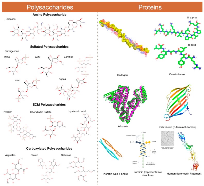
Biodegradable polymers are popularly being used in an increasing number of fields in the past few decades. The popularity and favorability of these materials are due to their remarkable properties, enabling a wide range of applications and market requirements to be met. Polymer biodegradable systems are a promising arena of research for targeted and site-specific controlled drug delivery, for developing artificial limbs, 3D porous scaffolds for cellular regeneration or tissue engineering and biosensing applications. Several natural polymers have been identified, blended, functionalized and applied for designing nanoscaffolds and drug carriers as a prerequisite for enumerable bionano technological applications. Apart from these, natural polymers have been well studied and are widely used in material science and industrial fields. The present review explains the prominent features of commonly used natural polymers (polysaccharides and proteins) in various nanomedical applications and reveals the current status of the polymer research in bionanotechnology and science sectors.
Keywords: biopolymers; drug delivery; nanoparticles; nanoscience; polymers; tissue engineering.
Conflict of interest statement
The authors declare no conflict of interest.
Figures


Similar articles
-
Critical Review of Biodegradable and Bioactive Polymer Composites for Bone Tissue Engineering and Drug Delivery Applications.Polymers (Basel). 2021 Aug 6;13(16):2623. doi: 10.3390/polym13162623. Polymers (Basel). 2021. PMID: 34451161 Free PMC article. Review.
-
Drug Release from Porous Matrixes based on Natural Polymers.Curr Pharm Biotechnol. 2017;18(9):721-729. doi: 10.2174/1389201018666171103141347. Curr Pharm Biotechnol. 2017. PMID: 29110601 Review.
-
Natural-origin polymers as carriers and scaffolds for biomolecules and cell delivery in tissue engineering applications.Adv Drug Deliv Rev. 2007 May 30;59(4-5):207-33. doi: 10.1016/j.addr.2007.03.012. Epub 2007 Apr 6. Adv Drug Deliv Rev. 2007. PMID: 17482309 Review.
-
Natural and Synthetic Polymers for Biomedical and Environmental Applications.Polymers (Basel). 2024 Apr 20;16(8):1159. doi: 10.3390/polym16081159. Polymers (Basel). 2024. PMID: 38675078 Free PMC article. Review.
-
Film forming microbial biopolymers for commercial applications--a review.Crit Rev Biotechnol. 2014 Dec;34(4):338-57. doi: 10.3109/07388551.2013.798254. Epub 2013 Aug 6. Crit Rev Biotechnol. 2014. PMID: 23919238 Review.
Cited by
-
Beyond Tissue replacement: The Emerging role of smart implants in healthcare.Mater Today Bio. 2023 Aug 29;22:100784. doi: 10.1016/j.mtbio.2023.100784. eCollection 2023 Oct. Mater Today Bio. 2023. PMID: 37731959 Free PMC article. Review.
-
Recent Advances in Polymeric Nanoparticle-Encapsulated Drugs against Intracellular Infections.Molecules. 2020 Aug 18;25(16):3760. doi: 10.3390/molecules25163760. Molecules. 2020. PMID: 32824757 Free PMC article. Review.
-
Biomechanical Properties and Biocompatibility of a Non-Absorbable Elastic Thread.J Funct Biomater. 2019 Nov 16;10(4):51. doi: 10.3390/jfb10040051. J Funct Biomater. 2019. PMID: 31744160 Free PMC article.
-
Nanoporous Microsponge Particles (NMP) of Polysaccharides as Universal Carriers for Biomolecules Delivery.Nanomaterials (Basel). 2020 May 31;10(6):1075. doi: 10.3390/nano10061075. Nanomaterials (Basel). 2020. PMID: 32486448 Free PMC article.
-
Modified Hyaluronic Acid-Laminin-Hydrogel as Luminal Filler for Clinically Approved Hollow Nerve Guides in a Rat Critical Defect Size Model.Int J Mol Sci. 2021 Jun 18;22(12):6554. doi: 10.3390/ijms22126554. Int J Mol Sci. 2021. PMID: 34207389 Free PMC article.
References
-
- Trindade T., Daniel-Da-Silva A.L. Biofunctional composites of polysaccharides containing inorganic nanoparticles. In: Hashim A., editor. Advances in Nanocomposite Technology. InTech; Rijeka, Croatia: 2011.
Publication types
LinkOut - more resources
Full Text Sources
Other Literature Sources
Miscellaneous
