. 2017 Aug 9;548(7666):175-182.
doi: 10.1038/nature23455.

The complete connectome of a learning and memory centre in an insect brain

Affiliations

The complete connectome of a learning and memory centre in an insect brain

Katharina Eichler et al. Nature. .

Abstract

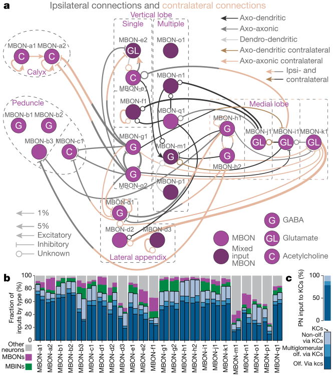
Associating stimuli with positive or negative reinforcement is essential for survival, but a complete wiring diagram of a higher-order circuit supporting associative memory has not been previously available. Here we reconstruct one such circuit at synaptic resolution, the Drosophila larval mushroom body. We find that most Kenyon cells integrate random combinations of inputs but that a subset receives stereotyped inputs from single projection neurons. This organization maximizes performance of a model output neuron on a stimulus discrimination task. We also report a novel canonical circuit in each mushroom body compartment with previously unidentified connections: reciprocal Kenyon cell to modulatory neuron connections, modulatory neuron to output neuron connections, and a surprisingly high number of recurrent connections between Kenyon cells. Stereotyped connections found between output neurons could enhance the selection of learned behaviours. The complete circuit map of the mushroom body should guide future functional studies of this learning and memory centre.


Figures

Extended Data Figure 1
Extended Data Figure 1. Connectivity of the larval APL neuron
a, Morphology of the right-hemisphere larval APL neuron. While APL dendrites are postsynaptic to the KCs in LA, LVL, IVL, and all medial lobe compartments, its axon is both post- and presynaptic to the MB calyx. Presynaptic sites in red and postsynaptic sites in blue. b, APL connectivity with KC types. Connections are displayed as fractions of input onto the receiving neurons. APL forms more axo-dendritic connections with multi-claw than with single-claw KCs. All mature KCs connect to APL on its dendrite as well as on its axon. c, Strength of synaptic connections between KCs and the APL neuron for single-claw and multi-claw KCs separately. While single-claw KCs have a higher synapse count connecting to the APL dendrites than multi-claw KCs, both groups of KCs project a similar number of synapses to the APL axon. In the calyx, APL makes more synapses onto multi-claw than onto single-claw KCs. In the lobes, single-claw KCs make more synapses with APL pre- than postsynaptically.
Extended Data Figure 2
Extended Data Figure 2. Input onto KC dendrites and KC–KC connections
a, Total synaptic input onto KC dendrites from PNs, the APL neuron, and calyx MBINs for single- and multi-claw KCs from both hemispheres. Insert: sum of the KC postsynaptic sites in the calyx as a function of the number of claws. Grey circles show individual KCs and black line shows the mean. Young KCs that have no claws or only form short branches in the calyx have few postsynaptic sites. Mature KCs forming one to six claws present a similar total amount of postsynaptic sites in the calyx. b, A PN bouton (blue) and its associated KC dendritic arborizations. Stars indicate all dendrites of the single-claw KC for this PN. c, Prototypical examples of KCs according to the structure of their dendrites, commonly known as ‘claws’. In first instar, we define each ‘claw’ (indicated by arrowheads) as a connection from a PN providing at least 10% of the postsynaptic sites of the KC dendritic arbor. This connectivity-based definition generally agrees with the count of physically separate claw-like dendritic branches that wrap around the axon terminals of the PNs. Single-claw KCs have not yet been described in the adult fly or other insects. d–g, Frequency of different numbers of postsynaptic KC-partners of KCs with at least two-synapse connections, plotted separately for three different types of KC. d, Postsynaptic partners of single-claw KCs. e, Postsynaptic partners of multi-claw KCs. f, Postsynaptic partners of young KCs. g, KC input onto KCs as a percentage of total KC input. For most KCs, more than 50% of their presynaptic partners are other KCs. h, Morphology of an example reconstructed KC (left) and example electron micrographs showing KC–KC synapses (right). Dendro-dendritic connections are in the calyx compartment (1 and 2) and axo-axonic connections are located in the peduncle (3), vertical lobe (4), and medial lobe (5).
Extended Data Figure 3
Extended Data Figure 3. Test of structure in PN-to-KC connectivity
a, We performed principal component analysis on the PN-to-KC connectivity matrices for the left and right hemispheres. The analysis was restricted to multi-claw KCs. The variance explained by each principal component in descending order is compared with that obtained from random models in which KCs sample PNs according to the individual PN connection probabilities (see Methods). Grey circles and bars denote mean and 95% confidence intervals for the variance explained by each principal component of the random connectivity matrices, while the black circles indicate the values obtained from data. Note a small deviation from the random model, probably because of the non-olfactory PNs. b, Same as a but restricted to connections between olfactory PNs and KCs. c, Same as b but combining the PN-to-KC connectivity for both hemispheres. The data are compared with a random model in which KCs in each hemisphere sample PNs randomly and independently (grey), and with a bilaterally symmetrical model in which the connectivity is random but duplicated across the two hemispheres (pink). d, To assess the ability of our method to identify structured connectivity, we also generated connectivity matrices in which a weak bias was added (blue). Networks were generated in which PNs were randomly assigned to one of two groups. For each KC, the probabilities of connecting to PNs belonging to one randomly chosen group were increased by 1%, while the probabilities for the other group were decreased by 1% (the baseline probabilities were on average approximately 5%). This procedure was performed independently for each KC and led to networks in which KCs preferentially sampled certain PNs. The data are inconsistent with this model, illustrating that biases of ∼ 1% connection probability can be identified using our methods. e, As an independent method to identify structure in the PN-to-KC wiring, we considered the distribution of olfactory PN overlaps for all KC pairs, defined as the number of olfactory PNs from which both KCs received input. This quantity specifically identified biases in the likelihood of KCs to sample similar inputs. As in b, no such structure was identified.
Extended Data Figure 4
Extended Data Figure 4. KC–KC clustering
Synaptic connectivity between KCs reveals two communities within the MB. a, Heat map representation of the KC–KC network adjacency matrix, sorted by community structure as discovered by the Louvain method, which identifies groups of KCs with more within-group connections than expected by chance. We denote the denser community on each side as ‘Group 1’ and the other community as ‘Group 2’. Number of cells in each group is shown in the column labels. b, The number of observed 2+ synapse connections between pairs of KCs within and between groups in the same side of the body, normalized by the total number of all possible such connections (L, left; R, right). c, Distribution of number of synapses per edge for connections within and between each group. Boxes indicate interquartile interval, whiskers the 95th percentile, cross indicates median, outliers shown. d, Claw distribution by group (both sides aggregated). Note that all single-claw KCs are in Group 1. e, Total number of anatomical input synapses onto Group 1 and Group 2 KCs, including from non-KC sources. Group 1 cells have significantly more inputs than Group 2 cells on each side (P <10-10, t-test with Bonferroni correction). f, Total number of anatomical output synapses from Group 1 and Group 2 KCs, including synapses onto non-KC targets. Group 1 cells have significantly more outputs than Group 2 cells on each side (P <10−10, t-test with Bonferroni correction). g, KC anatomy labelled by group for the right MB. Note that both groups of KCs come from each lineage cluster. Labelled spots indicate locations of cross-section views in h. h, Cross-sections of the two principal axon branches of KCs in the left (above) and right (below) MBs at the locations indicated in g. Orientations of each cross-section are arbitrary.
Extended Data Figure 5
Extended Data Figure 5. Neurons in the canonical circuit in every MB compartment
Neuronal morphology and connectivity of MBINs and MBONs participating in the canonical circuit motif of each MB compartment (MBINs in green and MBONs in magenta). Neurons connecting to the left-hemisphere MB are displayed in anterior view (right hemisphere has the same morphology data not shown). Locations of MBINs synapsing on MBONs in a given compartment are shown depending on their location (inside the MB neuropil, black; outside the MB, orange). MBIN axons and MBON dendrites tile the MB into 11 distinct compartments. For each compartment, the MBIN and MBON neurons and their connections as a fraction of total input to the receiving neuron are shown on the right. MBONs are diverse in the neurotransmitter that they release (see legend). All compartments present the canonical circuit motif except for SHA, which does not develop its DAN until later in larval life.
Extended Data Figure 6
Extended Data Figure 6. Fractions of postsynaptic inputs by cell type
a, Fractions of synaptic output from MBINs onto KCs, other MBINs, MBONs, and other neurons. Some MBINs show a high percentage of connections to MB neurons while others connect with less than 50% of their synapses to MB neurons. b, Number of MBIN–KC synapses in relation to the number of total synaptic outputs from MBIN axons, showing a positive correlation between presynaptic sites on the MBIN axon and synapses dedicated to KC population. c, Number of KC–MBON synapses in relation to the number of total synaptic inputs to MBON dendrites, showing a positive correlation between postsynaptic sites on the MBON dendrite and fraction of input from KCs.
Extended Data Figure 7
Extended Data Figure 7. MBIN presynaptic vesicle types
Examples of electron micrographs of MBIN presynaptic sites and vesicles. We found three types of vesicle: large dense-core, small dense-core, and small clear vesicles. Octopaminergic and dopaminergic neurons contain small clear vesicles in addition to other vesicle types. While OANs have all the same type of large dense-core vesicles, DANs show a variety of small dense-core vesicles. We found small dense-core vesicles in one-third of KCs. Some of these were single-claw, others were multi-claw, some received olfactory input, and others non-olfactory PN input. The largest number of dense-core vesicles was observed in the two thermosensory KCs. Scale bar, 500 nm in all panels.
Extended Data Figure 8
Extended Data Figure 8. Dense-core vesicles in OANs and DANs
a, Morphology of OAN-a1 and -a2 innervating the calyx of both MBs. b, Location of presynaptic sites (red) and dense-core vesicles (DCVs/black) along the axon of OAN-a1 and -a2. c, DCVs colour-coded by their distance to the closest presynaptic site on the axon of OAN-a1 and -a2. d, Distance (in micrometres) of DCVs to the closest presynaptic site sorted by the value for OAN-a1 and -a2. Most DCVs are within 2μm from a presynaptic site, just a few are further away and appear to be in transit. e, Presynaptic sites colour-coded by their distance to the closest DCV on the axon of OAN-a1 and -a2. f, Distance (in micrometres) of presynaptic sites to the closest DCV sorted by the value for OAN-a1 and -a2. Half of the presynaptic sites have a DCV associated within 20μm. Some presynaptic sites have no close DCV associated and are located in the dendrites of OAN-a1 and -a2 (data not shown). g, Morphology of DAN-i1 right innervating the upper toe of the MB medial lobe in both hemispheres with the location of presynaptic sites (red) and DCVs (black). h, Zoom-in onto the medial lobes from g. i, DCV colour-coded by their distance to the closest presynaptic site on the axon of DAN-i1 right. j, Presynaptic sites colour-coded by their distance to the closest DCV on the axon of DAN-i1 right. k, Distance (in micrometres) of DCVs to the closest presynaptic site sorted by the value for DAN-i1 right and left together. Some DCVs are further away from presynaptic sites than 10 μm; these DCVs are in the dendrites of the DAN (shown in g). And distance (in micrometres) of presynaptic sites to the closest DCV sorted by the value for DAN-i1 right and left together. Most of the presynaptic sites have a DCV associated within 1 μm.
Extended Data Figure 9
Extended Data Figure 9. KC-to-MBON synaptic connections
a, Percentage of mature KCs that are presynaptic to a given MBON. b, Frequency of the percentage of KCs presynaptic to MBONs (bin width is 10%). c, Frequency of the percentage of MBONs each KC connects to for single-claw and multi-claw KCs separately. All single-claw KCs connect with at least 75% of all MBONs present in their own hemisphere. d, Percentage of dendritic MBON inputs from individual KCs in the left brain hemisphere. KCs are ranked by their number of synapses to the MBON for each MBON separately (each line represents an MBON). Note the rank order of KCs is different for every MBON. Note also that a few MBONs receive very strong synaptic input from approximately 20 KCs and less than 3% from the remaining KCs, while most MBONs receive less than 4% of dendritic input from all KCs. e, Same as d, but for the right brain hemisphere. f, Effective strength of thermosensory input to MBONs. The thermosensory fraction is defined as the number of synapses received by an MBON from thermosensory KCs divided by 0.05 times the number of synapses received from all other KCs. The fraction thus represents the relative influence of an input that activates the thermosensory KCs compared with that of a typical stimulus that activates 5% of KCs.
Extended Data Figure 10
Extended Data Figure 10. MBIN-to-KC synaptic connections
a, Percentage of mature KCs that are postsynaptic to a given MBIN. On average, 68% of KCs in a compartment are postsynaptic to at least one MBIN of that compartment. b, Frequency of the percentage of KCs postsynaptic to MBINs (bin width is 10%). c, Frequency of the percentage of MBINs presynaptic to each KC for single-claw and multi-claw KCs separately. All single-claw KCs receive synaptic input from at least 50% of all MBINs present in their own hemisphere. d, Percentage of axonic outputs from MBINs connecting to individual KCs in the left brain hemisphere. KCs are ranked by their number of synapses they receive from an MBIN for each MBIN separately (each line represents an MBIN). Note the rank order of KCs is different for every MBIN. Note also that a few MBINs connect very strongly to approximately ten KCs and less than 2% to the remaining KCs, while most MBINs dedicate less than 2% of their axonic output to all KCs. e, Same as d, but for the right brain hemisphere.
Figure 1
Figure 1. Mushroom bodies of a first-instar Drosophila larva
a, KCs from an electron microscopy volume of the whole central nervous system. b, Associative learning in first-instar larvae: in a Petri dish, we presented an odour (cloud) and red light, either paired (left) or unpaired (right), and computed the performance index. Control larvae (attP2;UAS-CsChrimson) receiving paired stimuli did not learn, whereas larvae in which optogenetic activation of dopaminergic (PAM cluster) neurons (GMR58E02-GAL4;UAS-CsCrimson) was paired with odour showed robust appetitive learning (P < 0.0001). c, Diagram of the literature's MB circuitry model. PN relay sensory stimuli to KC dendrites. MBON dendrites and MBIN (DAN, OAN, and other) axons tile the parallel KC axons, defining compartments (coloured boxes). MBINs signal reward or punishment, and KCs synapse onto output neurons (MBONs). d, PN-to-KC connectivity, colour-coded by percentage of inputs on KC dendrites. Uniglomerular olfactory PNs (Olfactory PNs) and other PNs synapse onto single-claw or multi-claw KCs. Stars indicate KCs with identical input patterns on the left and right hemispheres. For the PNs on the right of the black vertical lines, the first 18 columns are left–right homologous PNs, the last column (left) and last five columns (right) are hemisphere-specific PNs. e, Number of KCs integrating inputs from uniglomerular olfactory PNs, multiglomerular olfactory PNs, non-olfactory PNs, or a mixture of these PN types. L, left; R, right. f, Earlier-born KCs join the lineage bundle closer to the neuropil surface than later-born ones, meaning older KCs present fewer claws than younger ones. Distances span from the point where the KC joins the bundle to the joining point of the KC nearest to the neuropil. Differences between all groups are significant (* * * P <0.0001; single-claw and multi-claw KC comparison P =0.0237).
Figure 2
Figure 2. KC connectivity reduces redundancy and optimizes stimulus discrimination
a, Distribution of KC claw numbers compared with random models. Random models have significantly fewer single-claw KCs (P < 10−5). Grey circles and lines denote mean and 95% confidence intervals. b, Classification error rate of a readout of the KC representation trained on a stimulus discrimination task. Observed connectivity (blue and orange) is compared with random models (grey) in which KCs have different distributions of average claw numbers. In a and b, grey circles and lines denote mean and 95% confidence intervals for random models. c, Average performance of models with purely random connectivity (grey) or random multi-claw plus non-random single-claw KCs (white). Standard error of the mean is smaller than the marks. d, Number of APL-to-KC synapses, which is correlated with claw number. e, Number of presynaptic KC-to-KC connections, which is inversely related to postsynaptic claw number. Dendro-dendritic (filled circles) or all synapses (open circles) are shown. In d and e, coloured circles and lines denote mean ± s.e.m. from the reconstructed data. f, Dimension of KC representation in models with only feedforward PN-to-KC connections (FF), with APL-mediated inhibition (FF + APL), or with inhibition and excitatory KC-to-KC connections (FF + APL + KC). Dimension is slightly reduced by dendritic KC-to-KC connections (right, filled bars) but strongly reduced by axonic KC-to-KC connections (open bars). Facilitatory axonic KC-to-KC connections (hatched bars) yield an intermediate reduction.
Figure 3
Figure 3. A canonical circuit in every MB compartment
a, Electron microscopy-reconstructed DAN/OAN/MBINs (green) and MBONs (magenta). b, Canonical circuit present in every MB compartment, with previously unknown KC synapses onto MBINs (DAN, OAN, and others), and from these onto MBONs. c, Example of an MBIN (green dot) synapse with a KC (white dot) and an MBON (magenta dot). The same KC is also presynaptic to the MBON in close proximity. Dense- and clear-core vesicles are present near the DAN presynaptic site. d, The connectivity matrix between KCs, DAN/OAN/MBINs, and MBONs of the right-hemisphere MB shows specific, compartment-centric synapses among cells types. Each entry represents the number of synapses from a row to a column (values are averaged for KCs). Note the absence of DAN in SHA (develops later in larval life). DAN/OAN/MBINs synapse only onto MBONs innervating their compartment, and axo-axonically onto same-compartment DAN/OAN/MBINs. Note multi-compartment MBONs in the vertical lobe and lateral appendix (LA). CA, calyx; IP, intermediate peduncle; LP, lower peduncle; LT, lower toe; UT, upper toe; IT, intermediate toe.
Figure 4
Figure 4. MBON inputs and circuits
a, MBONs are in the compartments innervated by their dendrites. Most connections among MBONs are axo-axonic, and fewer are axo-dendritic. Few MBONs avoid synapsing to others. Most inter-lobe connections are mediated by GABAergic (MBON-g1, -g2, -h1, -h2) and glutamatergic (MBON-i1, -j1, -k1) MBONs, potentially providing a substrate for lateral inhibition between compartments of opposite valence. b, Fraction of MBON inputs by neuron type. Left and right homologous MBONs are shown adjacent. Only some vertical lobe and lateral appendix MBONs get less than 80% of their input from MB neurons and less than 60% from KCs. Almost all multi-compartment MBONs (MBON-m1, -n1, -o1 and -p1) have a higher fraction of input from non-MB neurons than single-compartment MBONs. The fraction of inputs from PNs to MBONs via KCs is shown within the fraction of KC input (different shades of blue), computed by the product of the PN-to-KC and KC-to-MBON connections. Most MBONs receive a high fraction of olfactory input via KCs while few MBONs (-b3, -o1) get nearly half of their inputs via KCs from non-olfactory PNs. c, Percentages of different types of PN input to the KCs. While there are almost equal numbers of olfactory (olf.) and non-olfactory PNs synapsing onto KCs, non-olfactory PNs represent only about 12% of the inputs to KC dendrites.
Figure 5
Figure 5. Intra- and inter-compartment feedback: MBONs of one MB compartment synapse onto MBINs of the same or other compartments
Schematics of connections from output neurons (MBONs) onto input neurons (MBINs) within their own (feedback) or into other (feed-across) compartments. a, The output neuron MBON-e2 synapses onto the dendrites of OAN-e1 in its own compartment, UVL. b, Feedback among compartments of the same lobe suggests that the establishment of a memory in a compartment can affect the DANs of adjacent compartments. c, Feed-across motif from proximal MB compartments (calyx and intermediate peduncle) to a distal one (UVL). d, Feed-across motif from the vertical lobe to the medial lobe.

References

    1. Heisenberg M. Mushroom body memoir: from maps to models. Nat Rev Neurosci. 2003;4:266–275. - PubMed
    1. Farris SM. Are mushroom bodies cerebellum-like structures? Arthropod Struct Dev. 2011;40:368–379. - PubMed
    1. Menzel R. The honeybee as a model for understanding the basis of cognition. Nat Rev Neurosci. 2012;13:758–768. - PubMed
    1. Waddell S. Reinforcement signalling in Drosophila; dopamine does it all after all. Curr Opin Neurobiol. 2013;23:324–329. - PMC - PubMed
    1. Owald D, Waddell S. Olfactory learning skews mushroom body output pathways to steer behavioral choice in Drosophila. Curr Opin Neurobiol. 2015;35:178–184. - PMC - PubMed

Publication types