

Electron microscopy using the genetically encoded APEX2 tag in cultured mammalian cells
- PMID: 28796234
- PMCID: PMC5851282
- DOI: 10.1038/nprot.2017.065
Electron microscopy using the genetically encoded APEX2 tag in cultured mammalian cells
Abstract
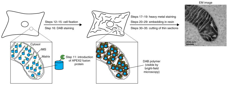
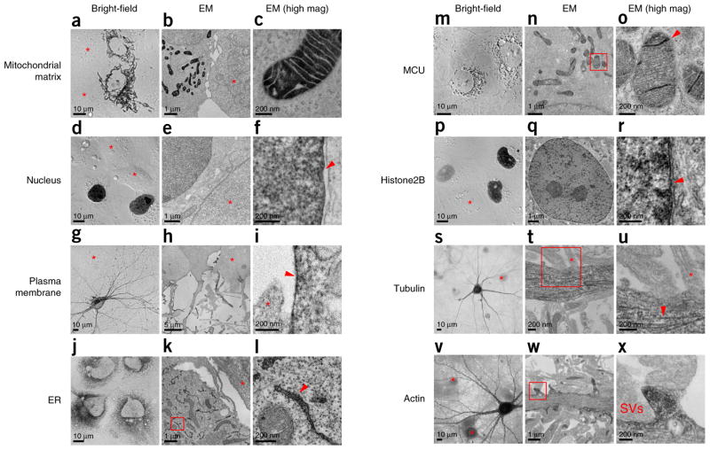
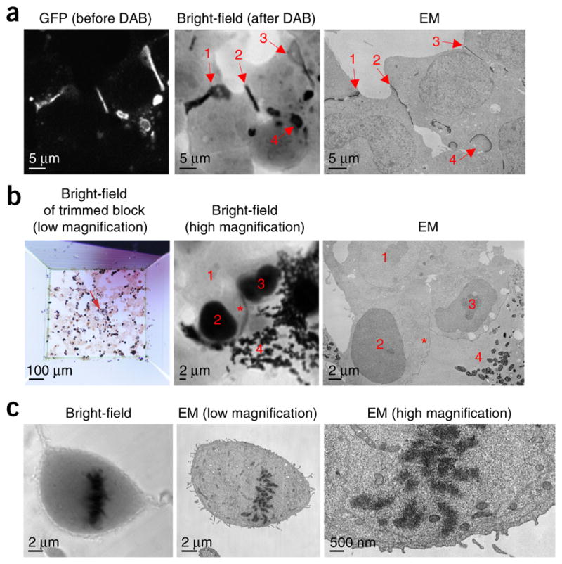
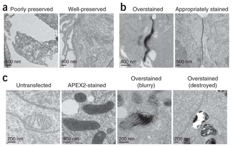
Electron microscopy (EM) is the premiere technique for high-resolution imaging of cellular ultrastructure. Unambiguous identification of specific proteins or cellular compartments in electron micrographs, however, remains challenging because of difficulties in delivering electron-dense contrast agents to specific subcellular targets within intact cells. We recently reported enhanced ascorbate peroxidase 2 (APEX2) as a broadly applicable genetic tag that generates EM contrast on a specific protein or subcellular compartment of interest. This protocol provides guidelines for designing and validating APEX2 fusion constructs, along with detailed instructions for cell culture, transfection, fixation, heavy-metal staining, embedding in resin, and EM imaging. Although this protocol focuses on EM in cultured mammalian cells, APEX2 is applicable to many cell types and contexts, including intact tissues and organisms, and is useful for numerous applications beyond EM, including live-cell proteomic mapping. This protocol, which describes procedures for sample preparation from cell monolayers and cell pellets, can be completed in 10 d, including time for APEX2 fusion construct validation, cell growth, and solidification of embedding resins. Notably, the only additional steps required relative to a standard EM sample preparation are cell transfection and a 2- to 45-min staining period with 3,3-diaminobenzidine (DAB) and hydrogen peroxide (H2O2).
Conflict of interest statement
Figures
References
-
- Giepmans BN, Adams SR, Ellisman MH, Tsien RY. The fluorescent toolbox for assessing protein location and function. Science. 2006;312:217–224. - PubMed
-
- Fernandez-Suarez M, Ting AY. Fluorescent probes for super-resolution imaging in living cells. Nat Rev Mol Cell Biol. 2008;9:929–943. - PubMed
-
- Xu K, Shim S-H, Zhuang X. In: Far-Field Optical Nanoscopy. Tinnefeld P, Eggeling C, Hell SW, editors. Springer; 2015. pp. 27–64.
-
- Eggeling C, Hell SW. In: Far-Field Optical Nanoscopy. Tinnefeld P, Eggeling C, Hell SW, editors. Springer; 2015. pp. 3–25.
-
- De Mey J, Moeremans M, Geuens G, Nuydens R, De Brabander M. High resolution light and electron microscopic localization of tubulin with the IGS (immuno gold staining) method. Cell Biol Int Rep. 1981;5:889–899. - PubMed
MeSH terms
Substances
Grants and funding
LinkOut - more resources
Full Text Sources
Other Literature Sources
Research Materials
