. 2017 Aug 10;8(8):e2985.
doi: 10.1038/cddis.2017.333.

P53-dependent downregulation of hTERT protein expression and telomerase activity induces senescence in lung cancer cells as a result of pterostilbene treatment

Affiliations

P53-dependent downregulation of hTERT protein expression and telomerase activity induces senescence in lung cancer cells as a result of pterostilbene treatment

Rong-Jane Chen et al. Cell Death Dis. .

Abstract

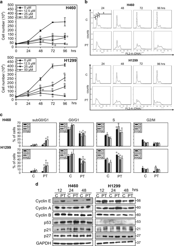
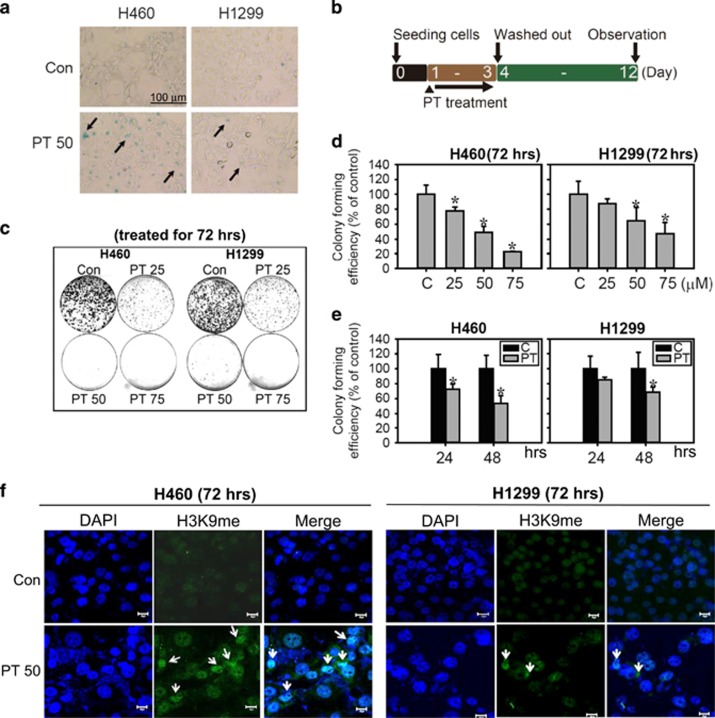
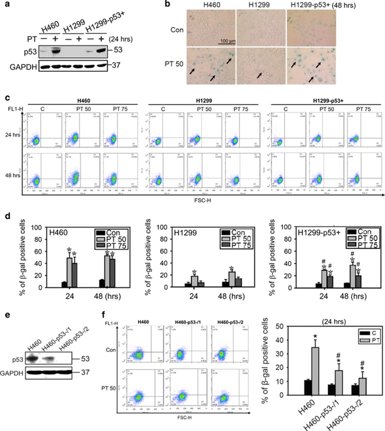
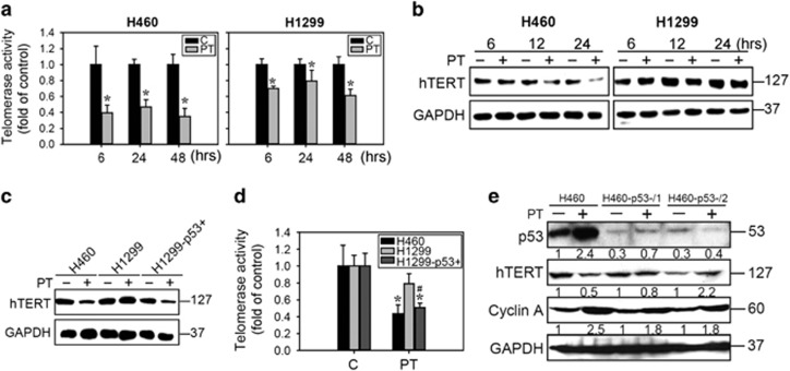
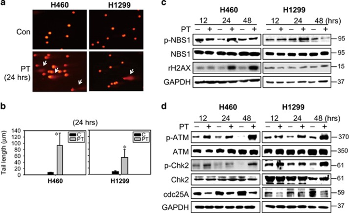
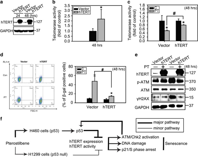
Cellular senescence is characterized by permanent cell cycle arrest, triggered by a variety of stresses, such as telomerase inhibition, and it is recognized as a tumor-suppressor mechanism. In recent years, telomerase has become an important therapeutic target in several cancers; inhibition of telomerase can induce senescence via the DNA damage response (DDR). Pterostilbene (PT), a dimethyl ether analog of resveratrol, possesses a variety of biological functions, including anticancer effects; however, the molecular mechanisms underlying these effects are not fully understood. In this study, we investigated the possible mechanisms of PT-induced senescence through telomerase inhibition in human non-small cell lung cancer cells and delineated the role of p53 in senescence. The results indicated that PT-induced senescence is characterized by a flattened morphology, positive staining for senescence-associated-β galactosidase activity, and the formation of senescence-associated heterochromatic foci. Telomerase activity and protein expression was significantly decreased in H460 (p53 wild type) cells compared with H1299 (p53 null) cells and p53 knockdown H460 cells (H460-p53-). A more detailed mechanistic study revealed that PT-induced senescence partially occurred via a p53-dependent mechanism, triggering inhibition of telomerase activity and protein expression, and leading to the DDR, S phase arrest and, finally, cellular senescence. This study is the first to explore the novel anticancer mechanism of PT senescence induction via the inhibition of telomerase in lung cancer cells.


Conflict of interest statement

The authors declare no conflict of interest.

Figures

Figure 1
Figure 1
Effects of PT on the growth inhibition and cell cycle arrest in lung cancer cells. (a) H460 and H11299 lung cancer cells were plated in six-well plates for 24 h and then treated with different concentrations of PT (0, 12.5, 25 and 50 μM) for 24, 48, 72 and 96 h. Cell numbers were counted daily by Trypan blue exclusion assay. Data represented the mean±S.E.M. of three independent experiments. *P<0.05 compared with the control group (0 μM). (b) H460 and H1299 were seeded in six-well plates and treated with 50 μM PT for the indicated times (24, 48, 72 and 96 h). Cells were collected and incubated with 40 μg/ml of PI for 15 min and subjected to flow cytometry analysis to examine the cell distribution at each phase of the cell cycle. Data are presented as representative graphs from three independent experiments. sub-G0/G1, G0/G1, S and G2/M phases are indicated as 1, 2, 3 and 4, respectively. (c) The percentage of cells in cell cycle phase was determined using FlowJo 7.6.1 software. Data represent the mean±S.E.M. (n=3, *P<0.05 compared with the control (d) H460 (left panel) and H1299 cells (right panel) were treated with 50 μM PT for indicated times then cell lysates were isolated and immunoblotted with anti-cyclin E, cyclin A, cyclin B, p53, p21 and p27 antibodies. Membranes were probed with an anti-GAPDH antibody to confirm equal loading of proteins. Representative data from one of three independent experiments are shown
Figure 2
Figure 2
PT-induced senescence in lung cancer cells. Senescence morphology of H460 and H1299 cells treated with 50 μM PT for 48 h. Cells were stained for β-gal. The representative images are shown with arrows indicating senescent morphology (a), bar: 100 μm. The experiment scheme for measuring loss of replication and regenerative potential (RP) was presented in (b). Cells were seeding overnight and then treated with or without 50 μM PT for 72 h. PT was then removed and cells could recover for additional 9 days (total period of 12 days). Then, colonies were stained with crystal violet (c). (d) Lung cancer cells were treated with 25, 50 or 75 μM PT for 72 h then PT was removed and the cells were allowed to recover for additional 9 days. (e) The colony-forming efficiency was also examined in lung cancer cells treated with 50 μM PT for 24 and 48 h and then cultivated in drug-free medium for additional 9 days. Data represent the mean±S.E.M. (n=3, *P<0.05 compared with the control). Y axis represents % decreases in the number of colonies relative to control. (f) Immunofluorescence analysis of the senescent heterochromatin foci stained with H3K9me3 (green) and with DAPI (blue) to visualize DNA in H460 and H1299 cells treated with PT (50 μM) for 72 h (Bar: 20 μm)
Figure 3
Figure 3
PT-induced senescence in lung cancer cells mediated by p53. (a) Protein expression of p53 in H460, H1299 and H1299-p53+ lung cancer cells after treated with 50 μM PT for 24 h. (b) Senescence morphology of H460 and H1299 cells treated with 50 μM PT for 48 h. Bar: 100 μm. (c) H460, H1299, and H1299-p53+ cells treated with 50 and 75 μM PT for 24 and 48 h and then incubated with C12FDG to detect SA-β gal activities by flow cytometry. X axis: FSC-H, Y axis: FL1-H. (d) The percentage of SA-β gal-positive cells detected by C12FDG staining is shown. Data represent the mean±S.E.M. of three independent experiments. *P<0.05, significantly higher than control group in different time course category. #P<0.05, significantly higher than H1299 groups. (e) Western blot analysis showed the expression of p53 in H460 cells and p53 stable knockdown cell lines (H460-p53-/1 and H460-p53-/2). (f) The percentage of SA-β gal-positive cells detected by C12FDG staining was shown in H460, H460-p53-/1 and H460-p53-/2 cells treated with 50 μM PT for 24 h. Data represent the mean±S.E.M. of three independent experiments. *P<0.05, significantly higher than control groups, #P<0.05, significantly lower than H460 PT-treated groups. X axis: FSC-H, Y axis: FL1-H
Figure 4
Figure 4
PT inhibited telomerase enzyme activity and protein expression in lung cancer cells. (a) H460 and H1299 cells were treated with 50 μM PT for the indicated times (6, 24 and 48 h) and telomerase enzyme activity were measured using a TRAPeze RT Telomerase Detection Kit. (b) Cell lysates extracted from H460 and H1299 cells treated with 50 μM PT for 6, 24 and 48 h were submitted to western blot analysis to detect the protein expression of hTERT. Equal loading was confirmed by GAPDH staining. (c) Western blot analysis for hTERT expression and (d) telomerase enzyme activity was measured by TRAPeze RT Telomerase Detection Kit in H460, H1299 and H1299-p53+ cells treated with 50 μM PT for 24 h. Data represented the mean±S.E.M. of three independent experiments. *P<0.05, compared with control groups. #P<0.05, significantly lower than H1299 groups. (e) Protein expression in H460, H460-p53-/1 and H460-p530-/2 cells treated with 50 μM PT for 24 h. The membrane was probed with anti-GAPDH to confirm equal loading of proteins. The number below each line indicates the relative intensity of protein expression compared with H460 control groups (p53) or each group without PT treatment (hTERT and cyclin A) (defined as 1)
Figure 5
Figure 5
PT-induced DNA damage in lung cancer cells. (a) Photomicrography of H460 and H1299 cells treated with 50 μM PT for 24 h and analyzed using comet assay. (b) DNA damage level expressed as tail length (μm) calculated using Komet 5.5 software (Kinetic Imaging Ltd., London, UK) after 24 h of 50 μM PT treatment in H460 and H1299 cells (mean±S.E.M., n=3, *P<0.05 compared with control groups). (c) H460 and H1299 cells were treated with 50 μM PT for 12, 24 and 48 h. Total-cell lysates were subjected to western blot analysis for (c) DNA damage proteins and (d) DNA sensor kinase (ATM), cell cycle checkpoint kinase (Chk2), and cell cycle regulatory protein (cdc25A). Membranes were probed with an anti-GAPDH antibody to confirm equal loading of proteins. Results are representative of three independent experiments
Figure 6
Figure 6
Exogenous telomerase expression rescued telomerase activity and decreased senescence induced by PT. (a) H460 cells were transiently transfected with either pcDNA-3.1 vector or pcDNA-3.1 hTERT-3HA plasmid for 24 and 48 h. Cell lysates were analyzed for the expression of hTERT by western blot analysis and (b) telomerase enzyme activity. (c) The telomerase enzyme activity of the vector or hTERT transfected H460 cells treated with 50 μM PT for 24 h were measured using a TRAPeze RT Telomerase Detection Kit. (mean±S.E.M., n=3, *P<0.05, significantly lower than control groups. #P<0.05, significantly higher than vector groups). (d) The SA β-gal activity of cells treated as in (b) was stained with C12FDG and analyzed by flow cytometry. X axis: FSC-H, Y axis: FL1-H. Data represented the mean±S.E.M. of three independent experiments. *P<0.05, compared with control groups. #P<0.05, significantly lower than vector groups. (e) Cells were treated as described in (b) and immunoblotting was performed with anti-hTERT, p-ATM, ATM, γH2AX, and GAPDH antibodies. Results are representative of three independent experiments. (f) Proposed model summarizing PT-induced senescence in lung cancer cells. In p53 wild-type H460 cells, PT inhibits hTERT enzyme activity and protein expression resulting in the subsequent induction of DNA damage, activation of ATM/Chk2 and p53, and S phase arrest. Activation of p53 positive feedback provokes hTERT downregulation, resulting in senescence in H460 cells. Interestingly, PT slightly inhibited hTERT enzyme activity resulting in less senescence in H1299 cells, suggesting that PT-induced senescence in lung cancer cells partially through p53-mediated hTERT inhibition

Similar articles

Cited by

References

    1. Rufini A, Tucci P, Celardo I, Melino G. Senescence and aging: the critical roles of p53. Oncogene 2013; 32: 5129–5143. - PubMed
    1. Bernhart E, Damm S, Heffeter P, Wintersperger A, Asslaber M, Frank S et al. Silencing of protein kinase D2 induces glioma cell senescence via p53-dependent and -independent pathways. Neuro Oncol 2014; 16: 933–945. - PMC - PubMed
    1. Luo H, Yang A, Schulte BA, Wargovich MJ, Wang GY. Resveratrol induces premature senescence in lung cancer cells via ROS-mediated DNA damage. PLoS ONE 2013; 8: e60065. - PMC - PubMed
    1. Kong Y, Cui H, Ramkumar C, Zhang H. Regulation of senescence in cancer and aging. J Aging Res 2011; 2011: 963172. - PMC - PubMed
    1. Kuilman T, Michaloglou C, Mooi WJ, Peeper DS. The essence of senescence. Genes Dev 2010; 24: 2463–2479. - PMC - PubMed

Publication types

MeSH terms