. 2017 Sep 20;28(9):2429-2439.
doi: 10.1021/acs.bioconjchem.7b00421. Epub 2017 Aug 10.

Multifunctional αvβ6 Integrin-Specific Peptide-Pt(IV) Conjugates for Cancer Cell Targeting

Affiliations

Multifunctional αvβ6 Integrin-Specific Peptide-Pt(IV) Conjugates for Cancer Cell Targeting

Anne C Conibear et al. Bioconjug Chem. .

Abstract

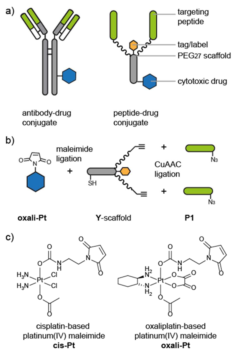
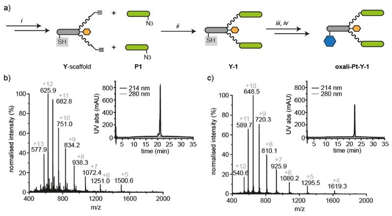
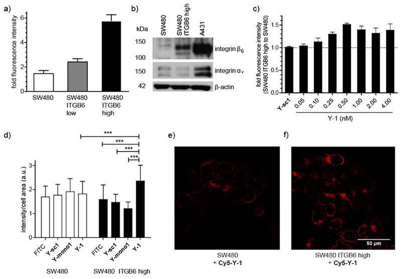
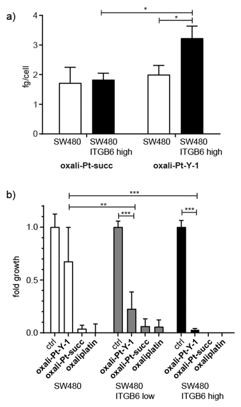
Increasing the specificity of cancer therapy, and thereby decreasing damage to normal cells, requires targeting to cancer-cell specific features. The αvβ6 integrin is a receptor involved in cell adhesion and is frequently up-regulated in cancer cells compared to normal cells. We have selected a peptide ligand reported to bind specifically to the β6 integrin and have synthesized a suite of multispecific molecules to explore the potential for targeting of cancer cells. A combination of solid-phase peptide synthesis and chemoselective ligations was used to synthesize multifunctional molecules composed of integrin-targeting peptides, cytotoxic platinum(IV) prodrugs, and fluorescent or affinity probes joined with flexible linkers. The modular synthesis approach facilitates the construction of peptide-drug conjugates with various valencies and properties in a convergent manner. The binding and specificity of the multifunctional peptide conjugates were investigated using a cell line transfected with the β6 integrin and fluorescence microscopy. This versatile and highly controlled approach to synthesizing labeled peptide-drug conjugates has the potential to target potent cytotoxic drugs specifically to cancer cells, reducing the doses required for effective treatment.


Conflict of interest statement

Notes

The authors declare no competing financial interest.

Figures

Figure 1
Figure 1. Design and assembly of peptide−drug conjugates.
(a) Comparison between a schematic antibody−drug conjugate and a schematic peptide−drug conjugate. The antigen-binding regions (green bar) of the antibody bind to specific receptors on cancer cells and are used to target the cytotoxic drug (blue hexagon) to the cancer cells. The peptide−drug conjugate is composed of targeting peptides (green) that bind to cancer cell receptors and a polyethylene glycol (PEG)−peptide scaffold that is conjugated to the cytotoxic drug. Chemical synthesis allows the inclusion of a label or tag (orange hexagon) and precise control over the targeting-moiety-to-drug ratio and location. (b) Modular synthesis and conjugation of the peptide−drug conjugate components. The maleimide-functionalized cytotoxic drug (oxali-Pt) is conjugated to a thiol on the peptide−PEG scaffold (Y-scaffold). The targeting peptides (P1) are conjugated to the scaffold via a copper-catalyzed azide−alkyne click (CuAAC) ligation. (c) Structures of maleimide-functionalized cis- and oxaliplatin-based platinum(IV) complexes cis-Pt and oxali-Pt.
Figure 2
Figure 2. Synthesis and characterization of peptide−drug conjugates.
(a) Solid-phase peptide synthesis (SPPS, i) was used to synthesize the azide-bearing targeting peptides (P1, green bar) and the alkyne-bearing peptide−PEG scaffold (Y) with the cysteine thiol protected (gray box). CuAAC ligation (ii) was used to conjugate the targeting peptides to the scaffold. The cysteine thiol was then deprotected (iii) and the maleimide-functionalized cytotoxic drug (oxali-Pt, blue hexagon) conjugated (iv). (b) ESI-MS and RP-HPLC traces of peptide−drug conjugate (oxali-Pt−Y-1) synthesized by modular synthesis (MWcalc 7499.5 and MWobs 7499.0). (c) ESI-MS and RP-HPLC traces of peptide−drug conjugate (oxali-Pt−Y-1) generated by synthesis of the combined Y-1 scaffold followed by maleimide ligation of oxali-Pt (MWcalc 6472.5 and MWobs 6471.3).
Figure 3
Figure 3. Integrin β6 transfection of SW480 colon carcinoma cells affects on the binding and uptake of targeted peptides.
(a) Integrin β6 expression of SW480 cells and transfected cells SW480 ITGB6 low (before sorting) and SW480 ITGB6 high (after sorting) given as fold fluorescence intensity relative to autofluorescence after binding of an antihuman integrin β6-allophycocyanin antibody and measured by flow cytometry in at least three independent experiments. (b) Integrin β6 and αv expression of SW480, SW480 ITGB6 high, and endogenously ITGB6-expressing A431 cells measured of total protein lysates by Western blot. β-actin levels were used as a loading control. Integrin β6 (MW 97 kDa) and integrin αv (MW 135−140 kDa). Full gels in Figure S2. (c) Binding of biotinylated Y-1 and nontargeting scrambled version biotinylated Y-sc1 (4 nM) to SW480 ITBG6 high compared to non-expressing SW480 cells. Fluorescence of peptide-binding avidin−FITC was measured by flow cytometry and normalized to samples without biotin-labeled peptide. Data is shown as the ratio between SW480 ITGB6 high compared to non-expressing SW480 cells, measured in three independent experiments. (d) Quantification of fluorescence intensity per cell area of confocal microscopy images of SW480 and SW480 ITGB6 high cells incubated with no peptide, 1 μM biotinylated Y-1, 1 μM biotinylated Y-mono-1, or 1 μM biotinylated Y-sc1. Before imaging, cells were fixed, permeabilized, and stained using avidin−FITC (1:200). At least 30 cells of each condition were quantified using ImageJ. (e, f) Confocal microscopy images representing (e) living SW480 and (f) SW480 ITGB6 high cells exposed to 0.5 μM Cy5−Y-1 for 10 min at 37 °C (scale bar: 50 μm). Values given in panels a, c, and d are the mean ± standard deviation. Significance was established using one-way ANOVA with Bonferroni’s multiple comparison test (***, p ≤ 0.001; **, p ≤ 0.01; and *, p ≤ 0.05).
Figure 4
Figure 4. Cellular uptake and cytotoxicity of peptide−drug conjugates in the SW480 model.
(a) Cellular uptake of oxali-Pt−Y-1 or oxali-Pt−succ by SW480 and SW480 ITGB6 high cells. Cells were incubated with 50 μM of either drug for 3 h before isolation and measurement by ICP-MS. The experiment was performed in triplicate. (b) Cytotoxicity of 10 μM oxali-Pt−Y-1, oxali-Pt−succ, and oxaliplatin after long-term exposure (14 days) to SW480, SW480 ITGB6 low, and SW480 ITGB6 high cells. Cell viability was measured by crystal violet staining from duplicates of three independent experiments. Values given in panels a and b are the mean ± standard deviation, and significances were established using one-way ANOVA with Bonferroni’s multiple comparison test (***, p ≤ 0.001; **, p ≤ 0.01, and *, p ≤ 0.05).

References

    1. Chari RV, Miller ML, Widdison WC. Antibody-drug conjugates: An emerging concept in cancer therapy. Angew Chem, Int Ed. 2014;53:3796–3827. - PubMed
    1. Casi G, Neri D. Antibody-drug conjugates: Basic concepts, examples and future perspectives. J Controlled Release. 2012;161:422–428. - PubMed
    1. Agarwal P, Bertozzi CR. Site-specific antibody–drug conjugates: The nexus of bioorthogonal chemistry, protein engineering, and drug development. Bioconjugate Chem. 2015;26:176–192. - PMC - PubMed
    1. Gilad Y, Firer M, Gellerman G. Recent innovations in peptide based targeted drug delivery to cancer cells. Biomedicines. 2016;4:11. - PMC - PubMed
    1. Wang Y, Cheetham AG, Angacian G, Su H, Xie L, Cui H. Peptide-drug conjugates as effective prodrug strategies for targeted delivery. Adv Drug Delivery Rev. 2017;110–111:112–126. - PMC - PubMed

Publication types

MeSH terms

LinkOut - more resources