. 2017 Aug 10;12(8):e0182153.
doi: 10.1371/journal.pone.0182153. eCollection 2017.

MyD88 contribution to ocular surface homeostasis

Affiliations

MyD88 contribution to ocular surface homeostasis

Rose Y Reins et al. PLoS One. .

Abstract

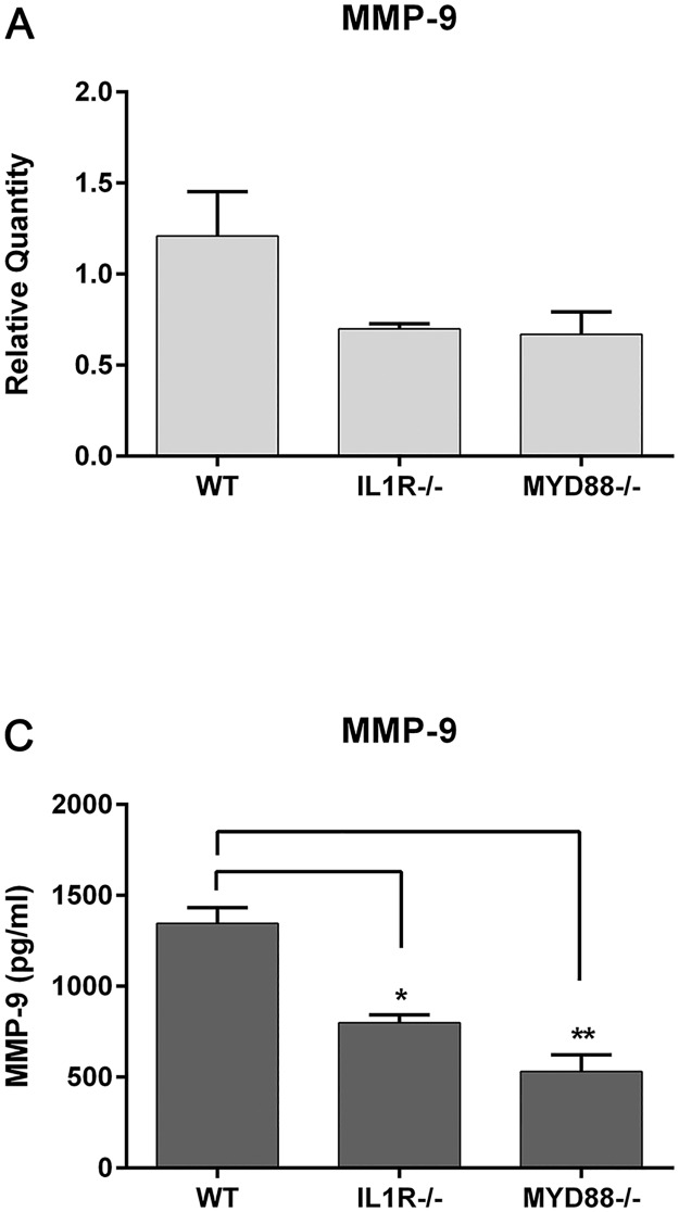
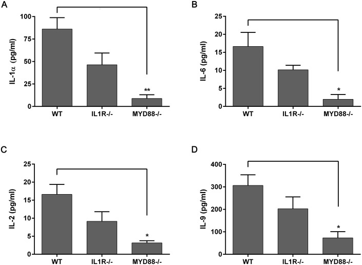
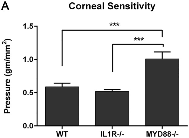
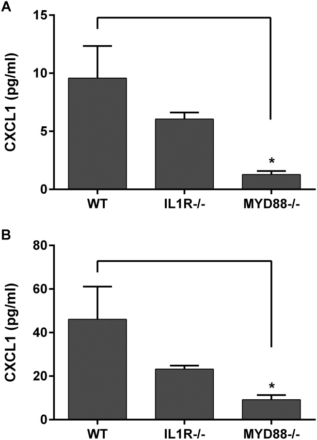
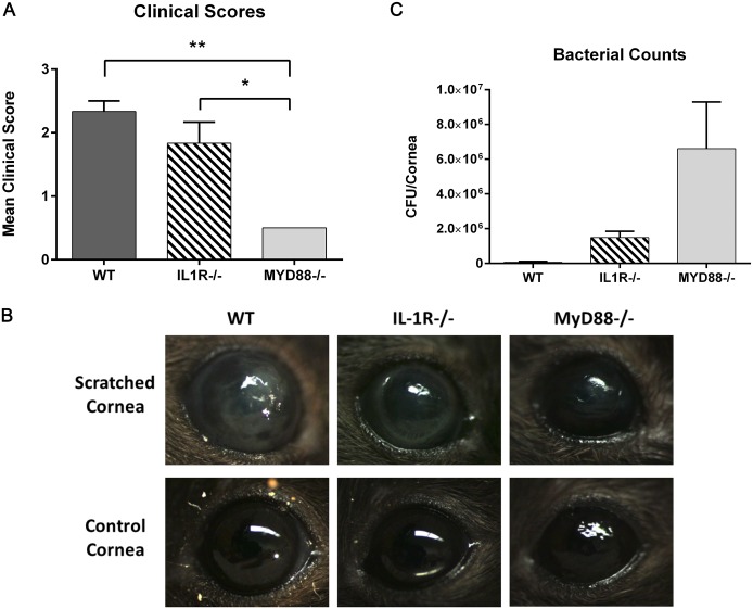
The cornea must maintain homeostasis, enabling rapid response to injury and microbial insult, to protect the eye from insult and infection. Toll-like receptors (TLRs) are critical to this innate immune response through the recognition and response to pathogens. Myeloid differentiation primary response (MyD88) is a key signaling molecule necessary for Toll-like receptor (TLR) and interleukin-1 receptor (IL-1R)-mediated immune defense and has been shown to be necessary for corneal defense during infection. Here, we examined the intrinsic role of TLR signaling in ocular surface tissues by determining baseline levels of inflammatory mediators, the response to mechanical stimuli, and corneal infection in MyD88-deficient mice (MyD88-/-). In addition, cytokine, chemokine, and matrix metalloproteinase (MMP) expression was determined in ocular surface cells exposed to a panel of TLR agonists. Compared to wild-type (WT) animals, MyD88-/- mice expressed lower MMP-9 levels in the cornea and conjunctiva. Corneal IL-1α, TNFα, and conjunctival IL-1α, IL-2, IL-6, and IL-9 levels were also significantly reduced. Additionally, CXCL1 and RANTES expression was lower in both MyD88-/- tissues compared to WT and IL-1R-/- mice. Interestingly, MyD88-/- mice had lower corneal sensitivities (1.01±0.31 gm/mm2) than both WT (0.59±0.16 gm/mm2) and IL-1R-/- (0.52±0.08 gm/mm2). Following Pseudomonas aeruginosa challenge, MyD88-/- mice had better clinical scores (0.5±0.0) compared to IL-1R-/- (1.5±0.6) and WT (2.3±0.3) animals, but had significantly more corneal bacterial isolates. However, no signs of infection were detected in inoculated uninjured corneas from either MyD88 or IL-1R-deficient mice. This work furthers our understanding of the importance of TLR signaling in corneal defense and immune homeostasis, showing that a lack of MyD88 may compromise the baseline innate response to insult.


Conflict of interest statement

Competing Interests: The authors have declared that no competing interests exist.

Figures

Fig 1
Fig 1. TLR activation increases levels of MMPs in human corneal and conjunctival cells.
Primary HCEC, hTCEpi, and primary HConjEpi were left untreated (control), or were treated with TLR agonists [TLR2/1 (PAM), TLR2 (HKLM), TLR4 (LPS), TLR5 (FLAG), TLR6/2 (FLS-1), TLR7 (IMQ), TLR8 (ssRNA), or TLR9 (ODN)] for 24 hours. Pro-MMP-9 (A), MMP-1 (B), and MMP-10 (C) expression was determined in cell supernatants by Luminex bead assay. Graphs represent mean ±SEM of 3 independent experiments (n = 3). Analysis was performed by ANOVA with Bonferroni’s test for multiple comparisons. p < *0.05, +0.01, #0.0001.
Fig 2
Fig 2. TLR agonist treatment induces cytokine expression in human corneal cells.
hTCEpi and primary HCEC were treated with TLR agonists [TLR2/1 (PAM), TLR2 (HKLM), TLR4 (LPS), TLR5 (FLAG), TLR6/2 (FLS-1), TLR7 (IMQ), TLR8 (ssRNA), or TLR9 (ODN)] for 24 hours and IL-8 (A, B) and IL-6 (C, D) expression was determined in cell supernatants by Luminex bead assay (hTCEpi) or ELISA (HCEC). Graphs represent mean ±SEM of 3 independent experiments (n = 3). Analysis was performed by ANOVA with Bonferroni’s test for multiple comparisons. p < *0.05, **0.01, ****0.0001.
Fig 3
Fig 3. MyD88-deficient mice have lower levels of MMP expression in the cornea and conjunctiva.
(A) MMP-9 expression was determined in untreated corneal lysates by RT-PCR. Graph represent mean ±SEM of 3 independent experiments with each sample pooled from 4 mice. (B) MMP-9 protein expression was quantitated in conjunctival homogenates by Luminex multiplex assay. Samples represent 10 pooled corneas per genotype. Graph represent mean ±SEM of 3 independent experiments (n = 3). Analysis was performed by ANOVA with Bonferroni’s test for multiple comparisons. p < *0.05, **0.01.
Fig 4
Fig 4. Lack of TLR signaling (MyD88-/-) results in decreased levels of cytokines in the cornea.
IL-1α (A, C) and TNFα (B, D) gene expression was determined in untreated corneal lysates by RT-PCR (top) and protein expression quantitated in corneal homogenates by Luminex multiplex assay (bottom), in wild-type (WT), IL-1R-/-, and MyD88-/- mice. Graphs represent mean ± SEM of 3 independent experiments with each sample pooled from 5 mice. Analysis was performed by ANOVA with Bonferroni’s test for multiple comparisons, with comparison to WT samples. p<*0.05, **0.01, nd = not determined.
Fig 5
Fig 5. MyD88-/- mice have decreased levels of cytokines in the conjunctiva.
IL-1α (A), IL-6 (B), IL-2 (C), and IL-9 (D) expression were determined in untreated conjunctival homogenates from wild-type (WT), IL-1R-/-, and MyD88-/- mice by Luminex multiplex assay. Data represent mean ± SEM of 3 independent experiments with each sample pooled from 5 mice. Analysis was performed by ANOVA with Bonferroni’s test for multiple comparisons, with comparison to WT samples. p<*0.05, **0.01.
Fig 6
Fig 6. MyD88-/- mice have decreased corneal sensitivity.
Corneal sensitivity was measured in untreated mice using a Cochet-Bonnet esthesiometer, with greater pressure (gm/mm2) indicating lower sensitivity. Graph represents mean ± SEM of 9 mice per genotype. Analysis was performed by ANOVA with Bonferroni’s test for multiple comparisons. p<***0.001.
Fig 7
Fig 7. Chemokine CXCL1 is decreased in MyD88-/- mice.
CXCL1 chemokine expression was determined in untreated corneal (top) and conjunctival (bottom) lysates from WT (C57), IL-1R-/-, and MyD88-/- mice by Luminex multiplex assay. Data represent mean ±SEM of 3 independent experiments with each sample pooled from 5 mice. Analysis was performed by ANOVA with Bonferroni’s test for multiple comparisons. p<*0.05.
Fig 8
Fig 8. MyD88-/- mice infected with PA were protected from inflammatory corneal infiltrates; however, more bacteria were isolated from MyD88-/- corneas compared to both WT and IL-1R-/-.
Corneas of WT (C57), IL-1R-/-, and MyD88-/- mice were scratched and infected with 1.0 × 10⁶ CFU GFP-PA01. (A-B) After 24 hours, eyes were imaged by slit lamp microscopy and graded based on severity of infection. Images are representative of 3 animals per group. Analysis was performed by ANOVA with Bonferroni’s test for multiple comparisons. p<*0.05, **0.01 (B) For bacterial counts, corneas were harvested 24 hours post-infection, homogenates plated, and incubated for 16 hours at 37°C. The number of colonies was counted and data represent 3 corneas per group.

References

    1. Takeda K, Kaisho T, Akira S. Toll-like receptors. Annu Rev Immunol. 2003;21: 335–376. doi: 10.1146/annurev.immunol.21.120601.141126 - DOI - PubMed
    1. Kawai T, Akira S. The role of pattern-recognition receptors in innate immunity: update on Toll-like receptors. Nat Immunol. 2010;11: 373–384. doi: 10.1038/ni.1863 - DOI - PubMed
    1. Kawasaki T, Kawai T. Toll-like receptor signaling pathways. Front Immunol. 2014;5: 461 doi: 10.3389/fimmu.2014.00461 - DOI - PMC - PubMed
    1. Redfern RL, Reins RY, McDermott AM. Toll-like receptor activation modulates antimicrobial peptide expression by ocular surface cells. Exp Eye Res. 2011;92: 209–220. doi: 10.1016/j.exer.2010.12.005 - DOI - PMC - PubMed
    1. Reins RY, Baidouri H, McDermott AM. Vitamin D Activation and Function in Human Corneal Epithelial Cells During TLR-Induced Inflammation. Invest Ophthalmol Vis Sci. 2015;56: 7715–7727. doi: 10.1167/iovs.15-17768 - DOI - PMC - PubMed

LinkOut - more resources