. 2017 Aug 10;12(8):e0182677.
doi: 10.1371/journal.pone.0182677. eCollection 2017.

Histological study on maturation, fertilization and the state of gonadal region following spawning in the model sea anemone, Nematostella vectensis

Affiliations

Histological study on maturation, fertilization and the state of gonadal region following spawning in the model sea anemone, Nematostella vectensis

Elizabeth Moiseeva et al. PLoS One. .

Abstract

The starlet sea-anemone Nematostella vectensis has emerged as a model organism in developmental biology. Still, our understanding of various biological features, including reproductive biology of this model species are in its infancy. Consequently, through histological sections, we study here key stages of the oogenesis (oocyte maturation/fertilization), as the state of the gonad region immediately after natural spawning. Germ cells develop in a secluded mesenterial gastrodermal zone, where the developing oocytes are surrounded by mucoid glandular cells and trophocytes (accessory cells). During vitellogenesis, the germinal vesicle in oocytes migrates towards the animal pole and the large polarized oocytes begin to mature, characterized by karyosphere formation. Then, the karyosphere breaks down, the chromosomes form the metaphase plate I and the eggs are extruded from the animal enclosed in a sticky, jelly-like mucoid mass, along with numerous nematosomes. Fertilization occurs externally at metaphase II via swimming sperm extruded by males during natural spawning. The polar bodies are ejected from the eggs and are situated within a narrow space between the egg's vitelline membrane and the adjacent edge of the jelly coat. The cortical reaction occurs only at the polar bodies' ejection site. Several spermatozoa can penetrate the same egg. Fertilization is accompanied by a strong ooplasmatic segregation. Immediately after spawning, the gonad region holds many previtellogenic and vitellogenic oocytes, though no oocytes with karyosphere. Above are the first histological descriptions for egg maturation, meiotic chromosome's status at fertilization, fertilization and the gonadal region's state following spawning, also documenting for the first time the ejection of the polar body.


Conflict of interest statement

Competing Interests: The authors have declared that no competing interests exist.

Figures

Fig 1
Fig 1. General view of the adult Nematostella vectensis.
A. Three main body parts are shown. Yellow arrowhead indicates the location of the gonadal region within the body; red arrowhead indicates the mesentery within the scapus. B. A schematic drawing of the gonadal region localization within the mesentery in transverse section. C. The egg masses with eggs and the nematosomes (arrowheads). eg, egg; jc, jelly coat; te, tentacles. Scale bars: A = 2.0 mm, C = 200 μm.
Fig 2
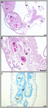
Fig 2. Longitudinal sections through a gravid Nematostella’s mesentery.
A. The upper part with tentacles (te), the scapulus (sl) and part of the scapus (sc) with the gonadal region (gr) (outlined) are seen. B. A higher magnification of the outlined zone in “A” reveals germ-cells groups with vitellogenic (ov) and previtellogenic (op) oocytes, and glandular cells surrounding them. Vitellogenic oocytes are strongly stained with azocarmine. PAF-Az stain. C. The germ-cells groups in the gonadal region, which contain selectively colored AB-positive glandular cells (gc) surrounding colorless oocytes (oo), can be seen. PAF-AB stain. gc, glandular cells; oo, colorless; oocyte; op, previtellogenic oocyte; ov, vitellogenic oocyte; sc, scapus; sl, scapulus; te, tentacles. Scale bars = 100 μm.
Fig 3
Fig 3. The gonadal region immediately after the extrusion of an egg mass.
A. A general view of the gonadal region in a cross section. Germ-cells groups are well visible. AHE stain; B. The longitudinal section of the gonadal region part. The gastric cavity (gs) is empty and clearly visible. Groups of germ cells at different stages of oogenesis and the glandular cells (gc) that surround them are seen within the gonadal region. PAF-AB-Az stain. C. Vitellogenic oocytes (ov) with an invagination of the oolemma (arrowhead). AHE stain. D. The part of the gonadal region seen in ‘B’ at a higher magnification. Note the bands of trophocytes (tr) between the glandular cells. PAF-AB-Az stain. E,F. Glandular cells show a reduced intensity of staining with reagent Schiff (PAS-AH stain in E) and alcian blue (AB-AHE stain in F) as compared to pre-spawning stage (Fig 4B and 4C; in both dyes, the fixation and staining periods were identical). In ‘E’ the vitellogenic oocytes with an invagination of the oolemma and trophonema (arrowheads) are seen. cl, cortical layer; gc, glandular cells; gs, gastric cavity; gv, germinal vesicle; op, previtellogenic oocyte; ov, vitellogenic oocyte; rm = retractor muscle; tr, trophocytes. Arrowheads in C show invagination of the oolemma; arrowheads in E show trophonema. Scale bars = 20 μm.
Fig 4
Fig 4. Oocytes of the different stages of oogenesis and the glandular cells surrounding the oocytes within the germ-cells groups.
A. The polarized previtellogenic oocyte, characterized by lamp-brush chromosomes (ch) inside the germinal vesicle, the oolemma’s invagination (iv) and the trophonema (arrowhead), which is well developed in the site of invagination. PAF-Az stain. B,C. Alcian blue-positive (B) and PAS-positive (C) glandular vacuolated cells (gc). The previtellogenic oocyte (op) (colorness in ‘B’) with nucleoli (nl) inside germinal vesicle (gv) and the vitellogenic oocytes (ov) are filled of the yolk granules (in ‘C’) can be seen. PAF-AB and PAS-AH stain, respectively. D. Histone H3 phosphorylation in prophase I meiosis chromatosomes in the germinal vesicle of a previtellogenic oocyte. E. The oocyte of the end of vitellogenesis. The germinal vesicle is located at the animal pole under the cortical layer (cl) of the ooplasm, and the ooplasm is filled with yolk granules and globules. The external edge of the cortical layer is visible”rough”. ch, chromosomes; cl, cortical layer; gc, glandular cells; gv, germinal vesicle; iv, invagination of the oolemma; nl, nucleoli; op, previtellogenic oocyte; ov, vitellogenic oocyte. Arrowhead shows trophonema (in A) AHE stain. Scale bars: A-D = 20 μm, E = 10 μm.
Fig 5
Fig 5. The karyosphere (ks) in maturating oocytes.
A. A general view of an oocyte with karyosphere. The yolk globules (gl) along the edge of the cortical layer (cl) and the space (sp) between the oolemma and the gastrodermal cells are seen. PAF-Az stain. B. Three karyosphere zones are visible: a relatively homogenous substance (ho), a denser, lumpy substance (arrowhead) and a ring-shaped zone. The chromosomes (cr) are inside the ring-shaped zone. AB-PAS stain. C. Condensed chromosomes (cr) are in the dense zone of the karyosphere. Note the space (sp) between the oolemma and the gastrodermal cells. DAPI stain. D. Chromosomes are in dense zone of the caryosphere during condensation. EtBr stain. cl, cortical layer; cr, chromosomes; dz, dense zone; gc, glandular cells; gl, yolk globules; ho, gomogenous zone; ks, karyosphere; op, previtellogenic oocyte; rz, ring-shaped zone; sp, space between the oolemma and gastrodermal cells. Arrowhead shows the dense zone of the karyosphere (in B). Scale bars = 20 μm.
Fig 6
Fig 6. Mature eggs during fertilization.
A. The metaphase II plate is located at the animal pole of the egg (arrowhead); AB-PAS-AH stain. B. The first polar body (arrowhead) that appeared in the space between the oolemma and edge of the jelly coat (jc). The segregated ooplasm (sg) is seen. AB-AHE stain. C. A general view of the animal pole during fertilization. The first polar body (arrowhead), the male pronucleus (mp), the female pronucleus (fp), the space (sp) between the oolemma and the edge of the jelly coat and the segregated ooplasm are seen. PAS-AB-AH stain. D. A higher magnification of the animal pole shown in ‘C’. The polar body (pb) is clearly seen in a state of division. E,F. The formation of two (E) and three polar bodies (F). The male pronucleus (arrowhead) is located in the peripheral zone of the egg (the coarse-grained yolk area) and the segregated ooplasm (in ‘E’) are clearly visible. AB-PAS-AH stain. G. Eggs located within the jelly coat. The ooplasmatic segregation is clearly visible. Inside the jelly coat are nematosomes (ns). AB-AHE stain. H. Numerous spermatozoa (sz) and one nematosome are seen inside the jelly coat. AHE stain. I. Some sperm heads (hs) are seen inside the eggs. They are visible as dark spots. AHE stain. J.K. The edge of the cortical layer (arrowheads) at the animal pole is characterized by a rough view. Fine granules are seen in the space between the oolemma and the internal edge of the jelly coat. In ‘J’ the female pronucleus is clearly seen, in ‘K’ the polar body is depicted. L. The edge of the cortical layer at the vegetative pole of the same egg is smooth (arrowhead). AB-AHE stain. cl, cortical layer; fp, female pronucleus; hs, sperm heads; jc, jelly coat; mp, male pronucleus; ns, nematosome; pb, polar body; sg, segregated ooplasm; sp, space between the oolemma and the edge of the jelly coat; sz, spermatozoa. Arrowhead in A shows metaphase II plate; arrowheads in B and C show the polar bodies; arrowheads in E and F show male pronuclei; yellow arrowheads in J and K illustrate a rough view of the cortical layer edge at animal pole and arrowhead in L shows a smooth view of the cortical layer at the vegetative pole in the same egg. Scale bars = 20 μm.

Similar articles

Cited by

References

    1. Williams RB. A redescription of the brackish water sea-anemone Nematostella vectensis Stephenson, with an appraisal of co-generic species. J Nat Hist 1975; 9: 51–64.
    1. Hand C, Uhlinger KR. The unique, widely distributed estuarine sea anemone, Nematostella vectensis Stephenson: a review new facts, and questions. Estuaries 1994; 17: 501–508.
    1. Daly M. A systematic revision of Edvardsiidae (Cnidaria, Anthozoa). Invert Biol 2002; 121: 212–225.
    1. Sullivan JC, Ryan JF, Watson JA, Webb J, Mullikin JC, Rokhsar D, et al. Stellabase: the Nematostella vectensis genomics database. Nucleic Acids Res 2006; 34: D495–499. doi: 10.1093/nar/gkj020 - DOI - PMC - PubMed
    1. Eckelbarger KJ, Hand C, Uhlinger KR. Ultrastructural features of the trophonema and oogenesis in the starlet sea anemone, Nematostella vectensis (Edvardsiidae). Invert Biol 2008; 127: 381–395.

LinkOut - more resources