. 2017 Aug 10;12(8):e0182102.
doi: 10.1371/journal.pone.0182102. eCollection 2017.

Repeat low-level blast exposure increases transient receptor potential vanilloid 1 (TRPV1) and endothelin-1 (ET-1) expression in the trigeminal ganglion

Affiliations

Repeat low-level blast exposure increases transient receptor potential vanilloid 1 (TRPV1) and endothelin-1 (ET-1) expression in the trigeminal ganglion

Elaine D Por et al. PLoS One. .

Abstract

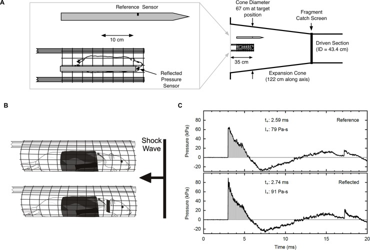
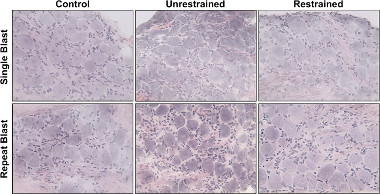
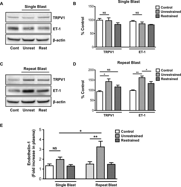
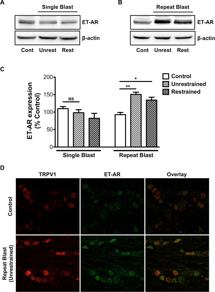
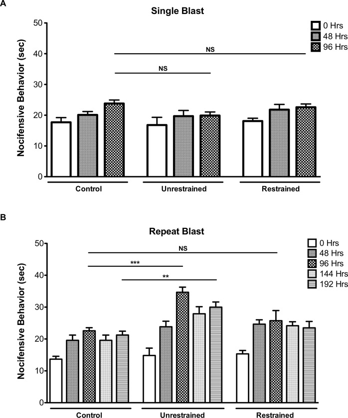
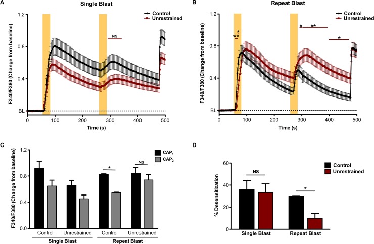
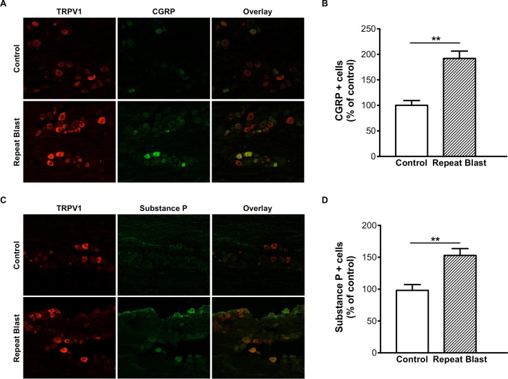
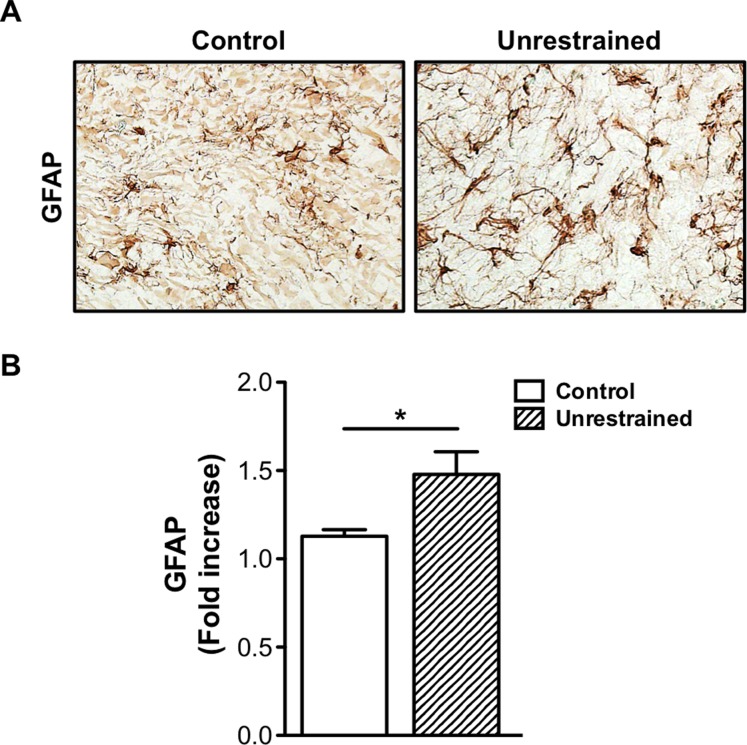
Blast-associated sensory and cognitive trauma sustained by military service members is an area of extensively studied research. Recent studies in our laboratory have revealed that low-level blast exposure increased expression of transient receptor potential vanilloid 1 (TRPV1) and endothelin-1 (ET-1), proteins well characterized for their role in mediating pain transmission, in the cornea. Determining the functional consequences of these alterations in protein expression is critical to understanding blast-related sensory trauma. Thus, the purpose of this study was to examine TRPV1 and ET-1 expression in ocular associated sensory tissues following primary and tertiary blast. A rodent model of blast injury was used in which anesthetized animals, unrestrained or restrained, received a single or repeat blast (73.8 ± 5.5 kPa) from a compressed air shock tube once or daily for five consecutive days, respectively. Behavioral and functional analyses were conducted to assess blast effects on nocifensive behavior and TRPV1 activity. Immunohistochemistry and Western Blot were also performed with trigeminal ganglia (TG) to determine TRPV1, ET-1 and glial fibrillary associated protein (GFAP) expression following blast. Increased TRPV1, ET-1 and GFAP were detected in the TG of animals exposed to repeat blast. Increased nocifensive responses were also observed in animals exposed to repeat, tertiary blast as compared to single blast and control. Moreover, decreased TRPV1 desensitization was observed in TG neurons exposed to repeat blast. Repeat, tertiary blast resulted in increased TRPV1, ET-1 and GFAP expression in the TG, enhanced nociception and decreased TRPV1 desensitization.


Conflict of interest statement

Competing Interests: The authors have declared that no competing interests exist.

Figures

Fig 1
Fig 1. Blast exposure experimental design.
(A) Top view of animal in expansion cone of shock tube with positions of reference and reflected sensors indicated. Diagram illustrates length of driven section and expansion cone of shock tube and cone diameter at target position. (B) Side view of animal with Kevlar vest in mesh holder. Top diagram illustrates unrestrained animal group and bottom diagram illustrates restrained animal group. (C) Representative shock wave produced by the USAISR shock tube as measured by the reflected pressure and overpressure sensors. Pressure represented in kilopascal (kPa). (t+ = time; duration of peak, I+ = impulse; area under peak).
Fig 2
Fig 2. H&E staining of trigeminal ganglion (TG) sections.
Frozen sections of TGs, from all animal treatment groups, were subjected to H&E staining to assess ganglion structure and anatomy. Staining revealed large sensory cell bodies surrounded by satellite cells. Results are representative images of all animal groups (40X magnification).
Fig 3
Fig 3. Repeat, tertiary low-level blast exposure increase TRPV1 and ET-1 expression in the trigeminal ganglion.
Western blot analysis was performed on TGs harvested from restrained or unrestrained animals exposed to either a single or repeat, low-level primary or tertiary blast. (A, C) TGs were homogenized and whole cell lysates (50 μg) were assessed for TRPV1 and ET-1 protein expression. (B, D) Quantification of TRPV1 and ET-1 expression in unrestrained and restrained animals following exposure to a single or repeat blast, respectively. Results are representative of four independent experiments. (E) Quantification of ET-1 expression in blood plasma from all control and blast treatment groups. Data represented as fold increase in ET-1 expression as compared to control. NS, not significant, *p<0.05 and **p<0.01, one-way ANOVA with Tukey post-hoc test.
Fig 4
Fig 4. Repeat blast exposure results in increased TRPV1 and ET-A co- expression.
Western blot and immunofluorescence analysis was performed on TGs harvested from animals that were exposed to either a single or repeat low-level primary or tertiary low-level blast. (A, B) TGs were homogenized and whole cell lysates (50 μg) were assessed for ET-A protein expression. (C) Quantification of ET-A expression in unrestrained and restrained animals following exposure to a single or repeat blast. (D) Immunofluorescence staining was completed on frozen sections of TGs subjected to repeat, tertiary blast. Tissues were subjected to staining with anti-TRPV1 (1:250) and anti-ET-A (1:250) antibodies and probed with Alexa Fluor 568 and 488 secondary antibodies, respectively. Representative images of an n = 4 for both control and repeat blast animals. NS, not significant, *p<0.05 and **p<0.01, one-way ANOVA with Tukey post-hoc test.
Fig 5
Fig 5. Repeat, tertiary blast exposure results in enhanced nocifensive behavior.
A dilute concentration of CAP (0.02%) was applied directly to the eye of unrestrained and restrained animals exposed to primary or tertiary low-level single (A) or repeat (B) blast. Eye wipe (nocifensive) responses were recorded for two minutes following application of CAP. Responses were counted as the amount of the time animal spent flinching and wiping the affected eye with either the paw or hind leg. Behavioral assessments were conducted at the indicated time points. Data are representative of 5–6 animals per treatment group. NS, not significant, **p<0.01and ***p<0.001, Repeated Measures ANOVA with Tukey post-hoc test.
Fig 6
Fig 6. Decreased TRPV1 desensitization in TG sensory neurons exposed to tertiary low-level blast.
TG were harvested from control animals and unrestrained animals exposed to a single or repeat, tertiary blast. Cumulative traces (A, B) and quantified responses (C) following initial and subsequent capsaicin (CAP) application, CAP1 and CAP2, respectively in KCl-sensitive TRPV1-expressing TG neurons. Shaded, yellow vertical bars denote application of CAP (50nM). (D) Quantified receptor desensitization of the indicated animal groups. NS, not significant, *p<0.05 and **p<0.01, two-way ANOVA with Bonferroni post-hoc test, n = 44–77 neurons/ group.
Fig 7
Fig 7. Increased CGRP and SP expression in TG sensory neurons exposed to repeat, tertiary blast.
Immunofluorescence analysis was performed on frozen TG sections harvested from control and repeat, tertiary (unrestrained) blast exposed animals. Tissues were subjected to staining with anti-TRPV1 (1:250), anti-CGRP (A) (1:250) and anti-SP (C) (1:250) antibodies and probed with the appropriate Alexa Fluor 568 and 488 secondary antibodies. (B, D) Quantification of CGRP- and SP-positive cells as percent of control animals, respectively. Paired t-test, **p<0.01.
Fig 8
Fig 8. Repeat low-level blast increases GFAP expression in the trigeminal ganglion.
(A) Immunohistochemical staining was performed on frozen TG sections harvested from control animals and unrestrained animals exposed to repeat, tertiary blast. Tissues were subjected to staining with anti-GFAP (1:1000) and then probed with the appropriate secondary biotinylated antibody. (B) Quantification of GFAP expression in TG homogenates from control and repeat, tertiary blast treatment groups. Data represented as fold increase in GFAP expression as compared to control. NS, not significant, *p<0.05, one-way ANOVA with Tukey post-hoc test.

References

    1. French LM. Military traumatic brain injury: an examination of important differences. Ann N Y Acad Sci. 2010;1208:38–45. doi: 10.1111/j.1749-6632.2010.05696.x . - DOI - PubMed
    1. Clark ME, Walker RL, Gironda RJ, Scholten JD. Comparison of pain and emotional symptoms in soldiers with polytrauma: unique aspects of blast exposure. Pain Med. 2009;10(3):447–55. doi: 10.1111/j.1526-4637.2009.00590.x . - DOI - PubMed
    1. Stratton KJ, Clark SL, Hawn SE, Amstadter AB, Cifu DX, Walker WC. Longitudinal interactions of pain and posttraumatic stress disorder symptoms in U.S. Military service members following blast exposure. J Pain. 2014;15(10):1023–32. doi: 10.1016/j.jpain.2014.07.002 ; PubMed Central PMCID: PMCPMC4213927. - DOI - PMC - PubMed
    1. Stratton KJ, Hawn SE, Amstadter AB, Cifu DX, Walker WC. Correlates of pain symptoms among Iraq and Afghanistan military personnel following combat-related blast exposure. J Rehabil Res Dev. 2014;51(8):1189–202. . - PMC - PubMed
    1. Bass CR, Panzer MB, Rafaels KA, Wood G, Shridharani J, Capehart B. Brain injuries from blast. Ann Biomed Eng. 2012;40(1):185–202. doi: 10.1007/s10439-011-0424-0 . - DOI - PubMed