. 2017 Aug 10;12(8):e0183189.
doi: 10.1371/journal.pone.0183189. eCollection 2017.

Uterotubal junction prevents chlamydial ascension via innate immunity

Affiliations

Uterotubal junction prevents chlamydial ascension via innate immunity

Yuyang Zhang et al. PLoS One. .

Abstract

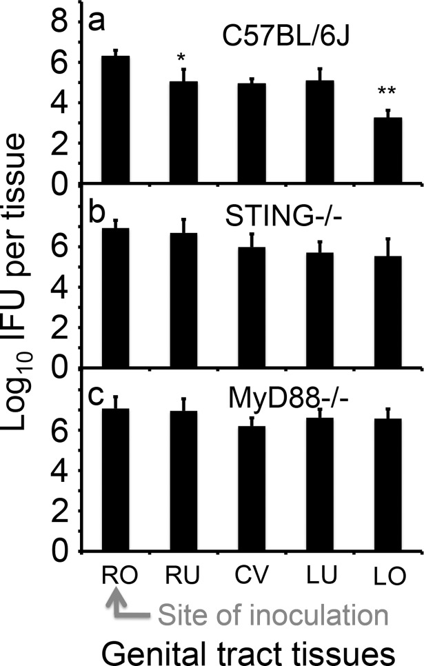
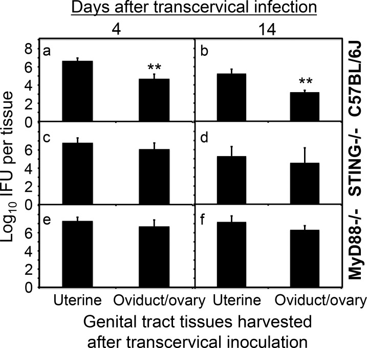
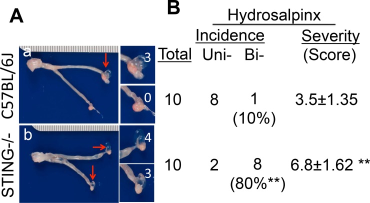
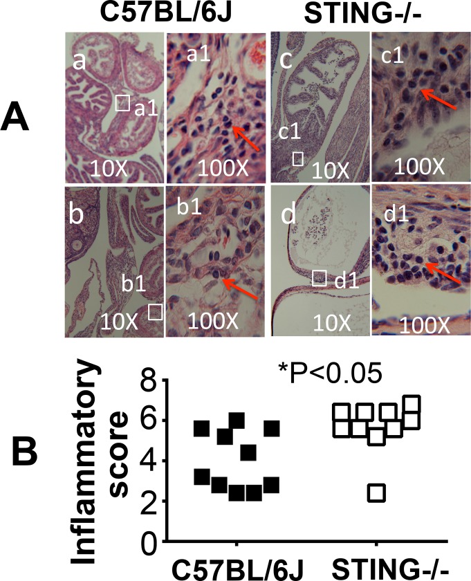
Ascension to the oviduct is necessary for Chlamydia to induce tubal infertility. Using the Chlamydia muridarum induction of hydrosalpinx mouse model, we have demonstrated a significant role of the uterotubal junction in preventing chlamydial ascending infection. First, delivery of C. muridarum to either side of the uterotubal junction resulted in significant reduction in live organisms from the tissues on the opposite sides. However, the recovery yields remained similar among different sections of the uterine horn. These observations suggest that the uterotubal junction may function as a barrier between the uterine horn and oviduct. Second, deficiency in innate immunity signaling pathways mediated by either MyD88 or STING significantly compromised the uterotubal junction barrier function, permitting C. muridarum to spread freely between uterine horn and oviduct. Finally, transcervical inoculation of C. muridarum led to significantly higher incidence of bilateral hydrosalpinges in the STING-deficient mice while the same inoculation mainly induced unilateral hydrosalpinx in the wild type mice, suggesting that the STING pathway-dependent uterotubal junction plays a significant role in preventing tubal pathology. Thus, we have demonstrated for the first time that the uterotubal junction is a functional barrier for preventing tubal infection by a sexually transmitted agent, providing the first in vivo evidence for detecting chlamydial infection by the STING pathway.


Conflict of interest statement

Competing Interests: The authors have declared that no competing interests exist.

Figures

Fig 1
Fig 1. Reduced spreading of Chlamydia muridarum from the oviduct/ovary into uterine horn after intrabursal injection.
The wild type C. muridarum organisms were intrabursally inoculated into the right oviduct/ovary of female C57BL/6J mice with 2 x 10⁵ inclusion forming units (IFUs) per mouse. At various time points post inoculation as indicated along the X-axis, groups of mice (n = 4 to 5) were sacrificed for harvesting the right oviduct/ovary (RO, solid bar) and right uterine horn (RU, open) respectively. The tissue samples were homogenized for titrating C. muridarum live organisms, and the titers were expressed as Log10 IFUs displayed along the Y-axis. Please note that titers of live organisms recovered from RO that received direct injection were always significantly higher than those from RU that is on the opposite side of the uterotubal junction (**p<0.01, *p<0.05, Wilcoxon), suggesting a functional barrier between the oviduct and uterine horn. The lower recovery during the first 24h is consistent with the concept that the inoculum needs time to replicate and to differentiate the replicating but non-infectious reticulate bodies into the infectious elementary bodies.
Fig 2
Fig 2. Comparing C. muridarum recoveries from 9 different sections of the mouse genital tract following inoculation into the right bursa.
A group of 5 C57BL/6J female mice were each inoculated with 2 x 10⁵ IFUs at the right bursa (as indicated with a red arrow) and 3 days after the inoculation, segments of the entire genital tract tissues were harvested as shown along with the X-axis, from the right oviduct/ovary (RO), right uterine horn distal region (RUd), middle (RUm) or the proximal region (RUp), cervico-vagina (CV), left side uterine horn proximal region (RUp), middle (LUm) and distal (RUd) to the left oviduct/ovary (LO). The tissues were homogenized for titrating infectious organisms as displayed in Log10 IFUs along the Y-Axis. Please note that significant differences in log10 IFUs were observed only between RO and RUd (*p<0.05, Wilcoxon) and between LUd and LO (**p<0.01, Wilcoxon) but not other adjacent tissue sections.
Fig 3
Fig 3. Comparing C. muridarum distribution in genital tract among three different mouse strains.
Groups of C57BL/6J (panel a, n = 5), Balb/c (b, n = 5) and CBA/1J (c, n = 5) female mice were each inoculated with 2 x 10⁵ IFUs at the right uterine horn distal region (RUd, as indicated with a red arrow) and 3 days after the inoculation, segments of the genital tract tissues were harvested as listed along the X-axis, from RO, RUd, RUm, RUp, CV, LUp, LUm, RUd to LO; tissue designations are indicated in Fig 2 legend The tissues were homogenized for titrating infectious organisms as displayed in Log10 IFUs along the Y-Axis. Please note that significant differences in log10 IFUs were observed between RO and RUd (**p<0.01, Wilcoxon) and between LUd and LO (**p<0.01, Wilcoxon) in all 3 mouse strains.
Fig 4
Fig 4. Evaluating the effect of MyD88 or STING pathways on the spread of C. muridarum in the genital tract following an inoculation to the right bursa.
Groups of C57BL/6J (panel a, n = 5), STING deficient (b, STING-/-, n = 5) and MyD88 deficient (c, MyD88-/-, n = 5) female mice were each inoculated with 2 x 10⁵ IFUs at the right bursa (RO, as indicated with a red arrow) and 3 days after the inoculation, segments of the genital tract tissues were harvested as listed along the X-axis, from RO, RU (entire right uterine horn), CV, LU (entire left uterine horn) to LO. The tissues were homogenized for titrating infectious organisms as displayed in Log10 IFUs along the Y-Axis. Please note that C57BL/6J mice displayed significant differences in log10 IFUs between RO and RU (*p<0.05, Wilcoxon) and between LU and LO (**p<0.01, Wilcoxon) respectively but neither STING-deficient nor MyD88-deficient mice were able to maintain the differences.
Fig 5
Fig 5. Evaluating the effect of MyD88 or STING pathways on C. muridarum ascending to the oviduct following a transcervical inoculation.
Groups of C57BL/6J (panels a & b, n = 5), STING-deficient (c & d, STNG-/-, n = 5) and MyD88-deficient (e & f, MyD88-/-, n = 5) female mice were each inoculated with 2 x 10⁵ IFUs transcervically and 4 (a, c & e) or 14 (b, d & f) days after the inoculation, the entire uterine horn (both right and left) and oviduct/ovary (both right and left) tissues from each mouse were harvested as listed along the X-axis. The tissues were homogenized for titrating infectious organisms as displayed in Log10 IFUs along the Y-Axis. Please note that C57BL/6J mice displayed significant differences in log10 IFUs between uterine and oviduct/ovary tissues (a & b, **p<0.01, Wilcoxon) but neither STING-deficient nor MyD88-deficient mice exhibited any significant differences.
Fig 6
Fig 6. Comparing C. muridarum recoveries from vaginal swabs of mice with or without deficiency in STING following a transcervical inoculation.
Groups of C57BL/6J (solid bar or, solid square, n = 10) and STING-deficient (open bar or open square, n = 10) female mice were each inoculated with 2 x 10⁵ IFUs transcervically and at various times after the inoculation as indicated along the X-axis, vaginal swabs were taken for titrating infectious organisms as displayed in Log10 IFUs (left) or % of mice with positive IFU (right) along the Y-Axis. Please note that there is no significant difference in either log10 IFUs (Wilcoxon) or % of mice with positive IFUs (Fisher’s Exact) between the wild type and STING-deficient mice (data obtained from two independent experiments), indicating that the STING-deficiency did not affect the descending of the C. muridarum organisms from the endocervcal compartments into the ectocervical and vaginal compartments or the replication of the C. muridarum organisms in these compartments.
Fig 7
Fig 7. Comparing gross pathology in the oviducts of mice with or without deficiency in STING following a transcervical inoculation.
Groups of C57BL/6J (n = 10) and STING-deficient (n = 10) female mice were each inoculated with 2 x 10⁵ IFUs transcervically as described in Fig 6 legend and 70 days after the inoculation, mice were sacrificed for evaluating gross pathology. (A) A representative image is shown from each group (A, panels a & b respectively). Hydrosalpinges were marked with red arrows while the severity of hydrosalpinx was scored based on the semi-quantitative scheme described in the materials and the scores were marked with white numbers. (B) Summary of both the hydrosalpinx incidence and severity. The incidence of mice with unilateral or bilateral hydrosalpinges was counted separately while the severity was scored based on total hydrosalpinges. Please note that only 10% the wild type C57BL/6J mice developed bilateral hydrosalpinx while 80% of the STING-deficient mice did so (**p<0.01, Fisher Exact). The STING-deficient mice also developed more severe hydrosalpinx than the wild type mice (**p<0.01, Wilcoxon).
Fig 8
Fig 8. Comparing inflammatory pathology in the oviducts of mice with or without deficiency in STING following a transcervical inoculation.
The genital tract tissues from the same mice as described in Fig 7 legend were subjected to H&E staining and evaluation of inflammatory infiltrates in both oviducts. (A) A representative H&E staining image is shown from each group (panels a & b from wild type C57BL/6J mice and c & d from STING-/- mice respectively under 10x objective lens). To visualize the infiltrating cells, examples of a 100x objective lens view are also provided as marked with a1 to d1. The inflammatory cells are marked with red arrows. (B) The H&E stained sections were semi-quantitatively scored based on the scheme described in the materials and method section, and the scores are summarized. Please note that the STING-deficient mice developed more severe inflammatory infiltration in the oviduct tissues (p<0.05, Wilcoxon).

Similar articles

Cited by

References

    1. Budrys NM, Gong S, Rodgers AK, Wang J, Louden C, Shain R, et al. Chlamydia trachomatis antigens recognized in women with tubal factor infertility, normal fertility, and acute infection. Obstetrics and gynecology. 2012;119(5):1009–16. doi: 10.1097/AOG.0b013e3182519326 ; PubMed Central PMCID: PMC4608258. - DOI - PMC - PubMed
    1. Rodgers AK, Budrys NM, Gong S, Wang J, Holden A, Schenken RS, et al. Genome-wide identification of Chlamydia trachomatis antigens associated with tubal factor infertility. Fertil Steril. 2011;96(3):715–21. Epub 2011/07/12. S0015-0282(11)00966-6 [pii] doi: 10.1016/j.fertnstert.2011.06.021 . - DOI - PMC - PubMed
    1. Rodgers AK, Wang J, Zhang Y, Holden A, Berryhill B, Budrys NM, et al. Association of tubal factor infertility with elevated antibodies to Chlamydia trachomatis caseinolytic protease P. American journal of obstetrics and gynecology. 2010;203(5):494 e7– e14. Epub 2010/07/21. S0002-9378(10)00705-2 [pii] doi: 10.1016/j.ajog.2010.06.005 . - DOI - PMC - PubMed
    1. Shah AA, Schripsema JH, Imtiaz MT, Sigar IM, Kasimos J, Matos PG, et al. Histopathologic changes related to fibrotic oviduct occlusion after genital tract infection of mice with Chlamydia muridarum. Sex Transm Dis. 2005;32(1):49–56. Epub 2004/12/23. 00007435-200501000-00008 [pii]. . - PubMed
    1. de la Maza LM, Pal S, Khamesipour A, Peterson EM. Intravaginal inoculation of mice with the Chlamydia trachomatis mouse pneumonitis biovar results in infertility. Infection and immunity. 1994;62:2094–7. - PMC - PubMed

MeSH terms