. 2017 Aug 10;12(8):e0183101.
doi: 10.1371/journal.pone.0183101. eCollection 2017.

A novel whole-bacterial enzyme linked-immunosorbant assay to quantify Chlamydia trachomatis specific antibodies reveals distinct differences between systemic and genital compartments

Affiliations

A novel whole-bacterial enzyme linked-immunosorbant assay to quantify Chlamydia trachomatis specific antibodies reveals distinct differences between systemic and genital compartments

Hannah L Albritton et al. PLoS One. .

Abstract

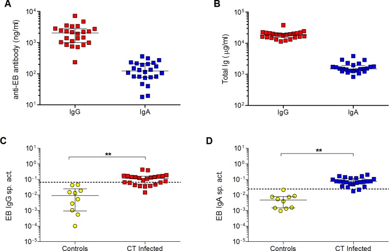
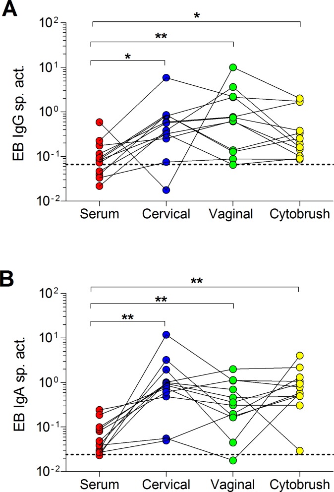
Chlamydia trachomatis (CT) is the leading sexually transmitted bacterial infection. The continued global burden of CT infection strongly predicates the need for a vaccine to supplement current chlamydial control programs. The correlates of protection against CT are currently unknown, but they must be carefully defined to guide vaccine design. The localized nature of chlamydial infection in columnar epithelial cells of the genital tract necessitates investigation of immunity at the site of infection. The purpose of this study was to develop a sensitive whole bacterial enzyme-linked immunosorbent assay (ELISA) to quantify and compare CT-specific IgG and IgA in sera and genital secretions from CT-infected women. To achieve this, elementary bodies (EBs) from two of the most common genital serovars (D and E) were attached to poly-L-lysine-coated microtiter plates with glutaraldehyde. EB attachment and integrity were verified by the presence of outer membrane antigens and the absence of bacterial cytoplasmic antigens. EB-specific IgG and IgA standards were developed by pooling sera with high titers of CT-specific antibodies from infected women. Serum, endocervical and vaginal secretions, and endocervical cytobrush specimens from CT-infected women were used to quantify CT-specific IgG and IgA which were then normalized to total IgG and IgA, respectively. Analyses of paired serum and genital samples revealed significantly higher proportions of EB-specific antibodies in genital secretions compared to sera. Cervical and vaginal secretions and cytobrush specimens had similar proportions of EB-specific antibodies, suggesting any one of these genital sampling techniques could be used to quantify CT-specific antibodies when appropriate normalization methodologies are implemented. Overall, these results illustrate the need to investigate genital tract CT antibody responses, and our assay provides a useful quantitative tool to assess natural immunity in defined clinical groups and CT vaccine trials.


Conflict of interest statement

Competing Interests: The authors have declared that no competing interests exist.

Figures

Fig 1
Fig 1. Poly-L-lysine coating of microtiter plates provides superior capture of EBs.
Plates coated with poly-L-lysine or PBS were treated with serial dilutions of a pooled EB mixture (CT serovars D and E) in duplicate, centrifuged, and then fixed with glutaraldehyde. The plates were then washed, blocked, and incubated with a saturating concentration (5.3 μg/ml) of mouse anti-CT LPS mAb. Mean absorbance values ± SD from one representative experiment of three are presented. Poly-L-lysine or PBS-coated plates identically treated with PBS served as negative controls. The concentration of EBs shown represents the total protein in the pooled EB mixture. A concentration of 4.5 μg/ml of pooled EBs and lysed EBs was selected for use in subsequent experiments.
Fig 2
Fig 2. Chlamydial antigens measured on captured EBs.
A total of 4.5 μg/ml of non-pooled or pooled EBs (red symbols) were fixed to poly-L-lysine-coated plates then incubated with duplicate serial dilutions of antibodies against chlamydial outer membrane proteins: (A) Non-pooled EBs of serovar D with mouse anti-CT LPS, (B) Non-pooled EBs of serovar E with mouse anti-CT LPS (C) Pooled EBs of serovars D and E with mouse anti-LPS (D) Pooled EBs of serovars D and E with mouse anti-MOMP D (E) Pooled EBs of serovars D and E with mouse anti-MOMP E and (F) Pooled EBs of serovars D and E with mouse anti-OmcA. Antibodies against cytoplasmic antigens of pooled EBs of serovars D and E: (G) Rabbit anti-Euo, (H) Mouse anti-Hsp60, and (I) Rabbit anti-Scc4. 4.5 μg/ml of lysed EBs of pooled serovars D and E were evaluated with (J) Rabbit anti-Euo (K) Mouse anti-Hsp60 and (L) Rabbit anti-Scc4. Blue symbols represent results obtained with irrelevant isotype-matched antibodies. Mean absorbance values ± SD from one representative experiment of three are shown.
Fig 3
Fig 3. Creation of EB IgG and IgA standards for antibody quantitation.
Sera from 20 CT-infected women were screened at a 1/1,000 dilution for (A) IgG antibodies and at a 1/50 dilution for (B) IgA antibodies that bound to pooled EBs. Shown is the mean absorbance ± SD for duplicate samples. The colored columns indicate sera that were subsequently pooled for use as the EB IgG or IgA standard and calibrated relative to total IgG and IgA standards, as described in the Methods. The dashed line represents the mean absorbance + 3 SD obtained with negative control IgG proteins at 10–20 μg/ml or IgA proteins at 20–40 μg/ml, concentrations approximately equivalent to those that would be present in serum diluted 1/1,000 (for IgG) or 1/50 (for IgA). (C) The EB IgG standard curve obtained after pooling the indicated sera is shown as mean ± SD for duplicate 2-fold serial dilutions, starting at a 1/1,000 dilution and a concentration of 19 ng/ml. (D) The EB IgA standard curve is similarly shown as mean ± SD for duplicate 2-fold dilutions starting at a 1/50 dilution and a concentration of 9.6 ng/ml. Using the EB IgG and IgA standards, (E) anti-EB IgG and (F) anti-EB IgA antibodies were measured in serum from 10 CT-infected women. Shown is the mean concentration ± SD obtained in two separate experiments. The inter-assay deviation for each sample is shown as the % CV (100% x SD/mean) above each column.
Fig 4
Fig 4. Quantification of EB-specific IgG and IgA in serum of CT-infected women.
(A) Concentrations of anti-EB IgG or IgA antibodies were measured in serum from 24 CT-infected women using the EB IgG or IgA standard. (B) Concentrations of total IgG and IgA in the sera were also measured by ELISA. The magnitude of systemic antibody responses to CT in infected women was determined by calculating the (C) EB IgG or (D) IgA specific activity (ng anti-EB IgG or IgA antibody per μg total IgG or IgA, respectively). The thresholds for significance in each assay are indicated by dashed lines representing the mean specific activity + 3 SD for negative controls (IgG or IgA myeloma proteins or human monoclonal antibodies to irrelevant antigens). Bars in all graphs denote medians and interquartile ranges. The EB specific activity in CT-infected women and controls was compared using the two-tailed Mann-Whitney rank sum test. **p < 0.01.
Fig 5
Fig 5. EB IgG and IgA specific activity in serum and genital tract secretions in CT-infected women.
(A) EB IgG and (B) IgA specific activity in matched serum, endocervical and vaginal secretions, and cytobrush samples were determined by dividing the anti-EB IgG or IgA antibody (in ng/ml) by the respective total IgG or IgA concentration (in μg/ml) in each sample. Dashed lines denote the thresholds for significance (mean specific activity + 3 SD of negative controls). *p < 0.05 and **p < 0.01 by two-tailed Wilcoxon matched pairs rank sum test.

References

    1. Newman L, Rowley J, Hoom SV, Wijesooriya NS, Unemo M, Low N, et al. Global estimates of the prevalence and incidence of four curable sexually transmitted infections in 2012 based on systematic review and global reporting. PLoS One. 2015;10(12). - PMC - PubMed
    1. Wyrick PB. Chlamydia trachomatis persistence in vitro: An overview. J Infect Dis. 2010;201 Suppl 2:S88–95. Epub 2010 May 28. - PMC - PubMed
    1. Peipert JF. Genital chlamydial infections. N Engl J Med. 2003;349:2424–30. doi: 10.1056/NEJMcp030542 - DOI - PubMed
    1. Darville T, Hiltke TJ. Pathogenesis of genital tract disease due to Chlamydia trachomatis. J Infect Dis. 2010;201 Suppl 2:S114–S25. - PMC - PubMed
    1. Golden MR, Schillinger JA, Markowitz LE, Louis ME. Duration of untreated genital infections with Chlamydia trachomatis: A review of the literature. Sex Transm Dis. 2000;27(6):329–37. - PubMed

MeSH terms