

Optical Coherence Tomography Angiography of the Peripapillary Retina in Primary Angle-Closure Glaucoma
- PMID: 28797550
- PMCID: PMC6524764
- DOI: 10.1016/j.ajo.2017.07.024
Optical Coherence Tomography Angiography of the Peripapillary Retina in Primary Angle-Closure Glaucoma
Abstract
Purpose: To measure the change of peripapillary retinal vessel density (VD) in eyes with a history of acute primary angle-closure glaucoma (PACG).
Design: Case-control study.
Methods: Twenty-one consecutive Chinese patients with history of unilateral acute PACG were enrolled. Eyes with acute PACG constituted the case group, while the contralateral eyes without attack constituted the control. All patients underwent ophthalmic examinations including best-corrected visual acuity, intraocular pressure, and visual field (VF). Spectral-domain optical coherence tomography (SD-OCT) was used to obtain both structural OCT and OCT angiography (OCTA). Structural OCT scans provided thickness measurements of the peripapillary retinal nerve fiber layer (RNFL) and macular ganglion cell complex (GCC). OCTA was used to measure all-plexus peripapillary retinal VD.
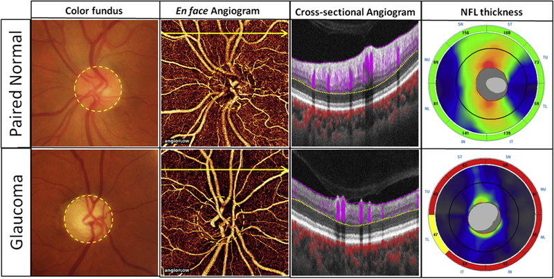
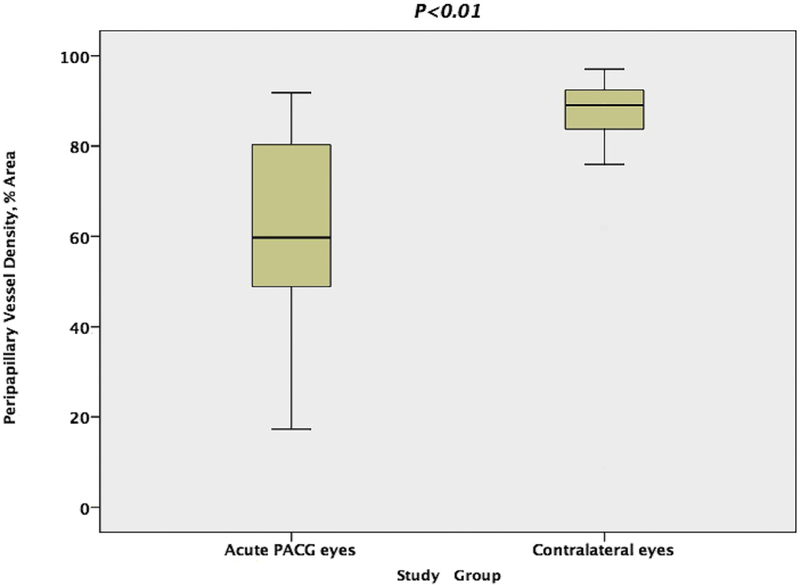
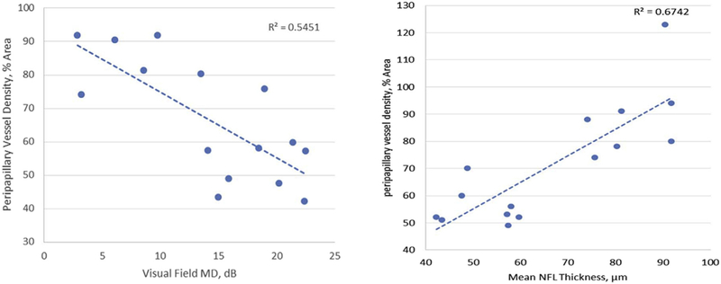
Results: In unaffected eyes, a dense microvascular network surrounded the disc on all-plexus retinal OCTA. The vascular network was visibly attenuated and focal capillary dropout was evident in acute PACG eyes. The peripapillary VD in acute PACG eyes was 66.6% ± 17.3% (mean ± standard deviation), which was significantly (P < .01) reduced compared to 87.2% ± 8.6% in the unaffected eyes. In acute PACG eyes, peripapillary retinal VD was positively correlated with RNFL and GCC thicknesses (P < .001 each) and negatively correlated with VF mean deviation (P = .002) and cup-to-disc ratio (P = .0064). In unaffected eyes, there were no correlations between peripapillary retinal VD and glaucoma-related parameters.
Conclusions: In acute PACG eyes, peripapillary retinal VD decreased significantly compared with the contralateral unaffected eyes. Peripapillary retinal VD was significantly correlated with other glaucomatous changes.
Copyright © 2017 Elsevier Inc. All rights reserved.
Figures
References
-
- Bourne RR, Stevens GA, White RA, et al. Causes of vision loss worldwide, 1990–2010: a systematic analysis. Lancet Glob Health 2013;1(6):e339–e349. - PubMed
-
- Tham YC, Li X, Wong TY, Quigley HA, Aung T, Cheng CY. Global prevalence of glaucoma and projections of glaucoma burden through 2040: a systematic review and meta-analysis. Ophthalmology 2014;121(11):2081–2090. - PubMed
-
- Yanagi M, Kawasaki R, Wang JJ, Wong TY, Crowston J, Kiuchi Y. Vascular risk factors in glaucoma: a review. Clin Exp Ophthalmol 2011;39(3):252–258. - PubMed
-
- Nongpiur ME, Ku JY, Aung T. Angle closure glaucoma: a mechanistic review. Curr Opin Ophthalmol 2011;22(2):96–101. - PubMed
-
- Costa VP, Harris A, Anderson D, et al. Ocular perfusion pressure in glaucoma. Acta Ophthalmol 2014;92(4):e252–e266. - PubMed
MeSH terms
Grants and funding
LinkOut - more resources
Full Text Sources
Other Literature Sources
