. 2017 Sep 4;214(9):2733-2758.
doi: 10.1084/jem.20161903. Epub 2017 Aug 10.

PTPN2 regulates T cell lineage commitment and αβ versus γδ specification

Affiliations

PTPN2 regulates T cell lineage commitment and αβ versus γδ specification

Florian Wiede et al. J Exp Med. .

Abstract

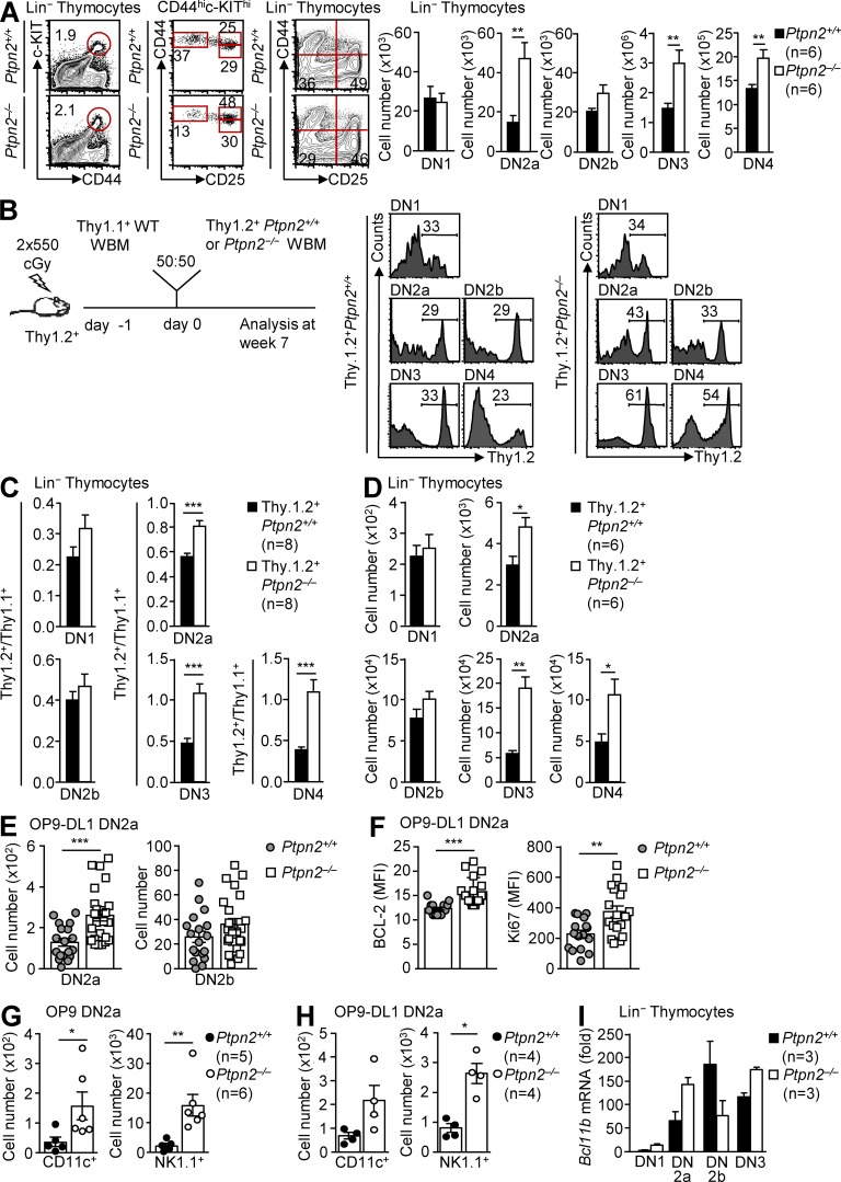
In the thymus, hematopoietic progenitors commit to the T cell lineage and undergo sequential differentiation to generate diverse T cell subsets, including major histocompatibility complex (MHC)-restricted αβ T cell receptor (TCR) T cells and non-MHC-restricted γδ TCR T cells. The factors controlling precursor commitment and their subsequent maturation and specification into αβ TCR versus γδ TCR T cells remain unclear. Here, we show that the tyrosine phosphatase PTPN2 attenuates STAT5 (signal transducer and activator of transcription 5) signaling to regulate T cell lineage commitment and SRC family kinase LCK and STAT5 signaling to regulate αβ TCR versus γδ TCR T cell development. Our findings identify PTPN2 as an important regulator of critical checkpoints that dictate the commitment of multipotent precursors to the T cell lineage and their subsequent maturation into αβ TCR or γδ TCR T cells.


Figures

Figure 1.
Figure 1.
PTPN2 deficiency influences thymocyte differentiation and increases STAT5 signaling. (A and B) Lineage (Lin) thymocytes from Ptpn2+/+ (C57BL/6) and Ptpn2/ (C57BL/6) mice (A) and poly (I:C)–treated Rosa26-eYFP;Ptpn2fl/fl (C57BL/6) or Mx1-Cre;Rosa26-eYFP;Ptpn2fl/fl (C57BL/6) mice (B) were stained with fluorochrome-conjugated antibodies against CD25, CD44, and c-KIT, and CD4/CD8 (DN) cell subsets were quantified by flow cytometry. (C) FACS-purified DN1 thymocytes from Ptpn2+/+ (C57BL/6) and Ptpn2/ (C57BL/6) mice were cultured for 10 d on OP9-DL1 stromal cells and stained for CD25, CD44, CD4, and CD8, and DN cell subsets were quantified by flow cytometry. (D, G, and H) Lin thymocytes from Ptpn2+/+ (C57BL/6) and Ptpn2/ (C57BL/6) mice were stained for CD25, CD44, and c-KIT and either intracellular p-(Y694) STAT5 (p-STAT5) (D), intracellular BCL-2 and MCL-1 (G), or the cell proliferation marker Ki67 (H) were determined in DN subsets by flow cytometry. (E) WBM cells from Ptpn2+/+ (C57BL/6) and Ptpn2/− (C57BL/6) mice were cultured on OP9-DL1 stromal cells for 9 d. Lin thymocytes were harvested and stained for CD25, CD44, and intracellular p-(Y694) STAT5 (p-STAT5). The mean fluorescence intensities (MFIs) for intracellular staining of p-STAT5 was determined by flow cytometry. (F) FACS-purified Linc-KIThiSCA-1hi BM cells from individual Ptpn2+/+ (C57BL/6) (n = 5) and Ptpn2/ (C57BL/6) (n = 5) mice were cultured on OP9-DL1 stromal cells for 9 d. DN2/3 thymocytes were harvested and stained for CD25 and CD44, and DN subsets were determined by flow cytometry. DN2/3 thymocyte lysates were resolved by SDS-PAGE and immunoblotted for p-STAT, STAT5, p-(Y1022/1023) JAK-1 (p-JAK1), JAK-1, PTPN2, and tubulin. Representative results (means ± SEM; Ptpn2+/+, n = 4–7; Ptpn2/, n = 4–9; Rosa26-eYFP;Ptpn2fl/fl, n = 4; Mx1-Cre;Rosa26-eYFP;Ptpn2fl/fl, n = 4) and representative cytometry profiles (C, D, and F) and immunoblots (F) from at least two independent experiments are shown. In F, each lane represents the cell lysate from one individual mouse. Significance was determined using two-tailed Mann-Whitney U test; *, P < 0.05; **, P < 0.01; ***, P < 0.001.
Figure 2.
Figure 2.
PTPN2 deficiency represses T cell lineage commitment. (A) Lin thymocytes from Ptpn2+/+ (C57BL/6) and Ptpn2/ (C57BL/6) mice were stained for CD25, CD44, and c-KIT, and DN cell subsets were quantified by flow cytometry. (B–D) Equal numbers of donor WBM cells (2 × 10⁶) from Thy1.2+Ptpn2+/+ (BALB/c) or Thy1.2+Ptpn2/ (BALB/c) mice and congenic Thy1.1+ (BALB/c) competitor cells were transferred into lethally irradiated (2 × 550 cGy) BALB/c recipient animals. Donor cell contribution in the thymus was assessed at 7 wk after transplantation. (B) Representative FACS profiles showing percent donor (Thy1.2)-derived DN thymocyte subset reconstitution. (C) Donor Thy1.2+Ptpn2+/+ (BALB/c) or Thy1.2+Ptpn2/ (BALB/c) cells and Thy1.1+ competitor (BALB/c) DN subset ratios. (D) Absolute numbers of donor Thy1.2+Ptpn2+/+ (BALB/c) versus Thy1.2+Ptpn2/ (BALB/c) DN subsets. (E and F) DN2a thymocytes from Ptpn2+/+ (C57BL/6) and Ptpn2/ (C57BL/6) mice were single-cell sorted, cultured on OP9-DL1 stromal cells for 72 h, and then stained with fluorochrome-conjugated antibodies against CD25 and c-KIT. DN2a and DN2b cells were quantified by flow cytometry (E), and intracellular BCL-2 and Ki67 MFIs in DN2 cells were determined (F). (G and H) 102 DN2a thymocytes from Ptpn2+/+ (C57BL/6) and Ptpn2/ (C57BL/6) mice were FACS purified, cultured on OP9 (G) or OP9-DL1 (H) stromal cells for 5 d, and then stained with fluorochrome-conjugated antibodies against Lin markers, CD11c, and NK1.1. Cell subsets were quantified by flow cytometry. (I) Bcl11b mRNA levels in Ptpn2+/+ (C57BL/6) and Ptpn2/ (C57BL/6) DN thymocyte subsets were assessed by quantitative RT-PCR. Representative results (means ± SEM; Ptpn2+/+, n = 3–6; Ptpn2/, n = 3–6; Thy1.2+.Ptpn2+/+, n = 6–8; and Thy1.2+.Ptpn2−/−, n = 6–8) and representative cytometry profiles (A and B) from at least three independent experiments are shown. In E and F, pooled data from three independent experiments are shown. In A, C, D, G, and H, significance was determined using two-tailed Mann-Whitney U test; *, P < 0.05; **, P < 0.01; ***, P < 0.001. In E and F, significance was determined using two-tailed Student’s t test. **, P < 0.01; ***, P < 0.001.
Figure 3.
Figure 3.
STAT5 heterozygosity versus JAK PTK inhibition corrects DN thymocyte development associated with PTPN2 deficiency. (A) Lin thymocytes from poly (I:C)–treated Rosa26-YFP;Ptpn2fl/fl (C57BL/6), Mx1-Cre;Rosa26-eYFP;Ptpn2fl/fl;Stat5+/ (C57BL/6), Mx1-Cre;Rosa26-eYFP;Ptpn2fl/fl (C57BL/6), and Rosa26-eYFP;Ptpn2fl/fl;Stat5+/ (C57BL/6) mice were stained for CD25, CD44, and c-KIT, and DN cell subsets were quantified by flow cytometry. (B) WBM cells from Rosa26-eYFP;Ptpn2fl/fl (C57BL/6), Mx1-Cre;Rosa26-eYFP;Ptpn2fl/fl(C57BL/6);Stat5+/ (C57BL/6), and Mx1-Cre;Rosa26-eYFP;Ptpn2fl/fl (C57BL/6) mice were cultured on OP9-DL1 stromal cells. Lin thymocytes were harvested after 10 d and stained for CD25 and CD44, and DN cell subsets were quantified by flow cytometry. (C) WBM cells from Ptpn2+/+ (C57BL/6) and Ptpn2/ (C57BL/6) mice were cultured on OP9-DL1 stromal cells in the presence of the JAK PTK inhibitor CMP6 or vehicle control (DMSO). DN2 and DN3 cell numbers were quantified by flow cytometry. Representative results (means ± SEM; Ptpn2+/+, n = 4; Ptpn2/, n = 4; Rosa26-eYFP;Ptpn2fl/fl, n = 5–6; Mx1-Cre;Rosa26-eYFP;Ptpn2fl/fl, n = 5–7; Mx1-Cre;Rosa26-eYFP;Ptpn2fl/fl;STAT5fl/+, n = 5–8; and Rosa26-eYFP;Ptpn2fl/fl;STAT5fl/+, n = 7) from at least three independent experiments are shown. Significance was determined using two-tailed Mann-Whitney U test. *, P < 0.05; **, P < 0.01; ***, P < 0.001.
Figure 4.
Figure 4.
PTPN2 overexpression represses DN thymocyte development. (A–C) 5 × 10⁵ WBM cells from C57BL/6 mice were transduced with control mCherry-expressing retrovirus (MSCV) or retroviruses expressing mCherry plus WT PTPN2, nuclear-restricted GFP-PTPN2 (A and B), or catalytically inactive PTPN2-R222M (C) and were transplanted into lethally irradiated (2 × 550 cGy) C57BL/6 hosts. DN thymocyte development was assessed after 7 wk. (A) Intracellular PTPN2 levels (MFI) in Lin mCherry+ DN thymocytes from mice reconstituted with WBM transduced with MSCV control–, WT PTPN2–, or GFP-PTPN2–expressing retroviruses were determined by flow cytometry. (B and C) Relative mCherry+ DN1–4 cells from mice reconstituted with WBM cells infected with control MSCV or MSCV encoding WT PTPN2 or GFP-PTPN2 (B), or mice reconstituted with WBM cells infected with control MSCV or MSCV encoding PTPN2-R222M (C). Representative results (means ± SEM; MSCV, n = 4–8; PTPN2, n = 4–6; PTPN2-GFP, n = 4–6; and PTPN2-R222M, n = 7) and representative cytometry profiles from at least two independent experiments are shown. Significance was determined using two-tailed Mann-Whitney U test. *, P < 0.05; **, P < 0.01.
Figure 5.
Figure 5.
PTPN2 regulates STAT5-dependent T cell lineage commitment. (A) Lin thymocytes from poly (I:C)–treated Rosa26-YFP;Ptpn2fl/fl (C57BL/6), Mx1-Cre;Rosa26-eYFP;Ptpn2fl/fl (C57BL/6), and Mx1-Cre;Rosa26-eYFP;Ptpn2fl/fl;Stat5fl/+ (C57BL/6) mice were stained for CD25, CD44, and c-KIT, and DN2a and DN2b cell subsets were quantified by flow cytometry. (B and C) FACS-purified Lin DN2a thymocytes from poly (I:C)–treated Rosa26-eYFP;Ptpn2fl/fl (C57BL/6), Mx1-Cre;Rosa26-eYFP;Ptpn2fl/fl;Stat5+/ (C57BL/6), and Mx1-Cre;Rosa26-eYFP;Ptpn2fl/fl (C57BL/6) mice were cultured on OP9-DL1 stromal cells for 3 d (B) or OP9 cells for 5 d (C) and then stained for c-KIT, CD44, and CD25 or lineage markers CD11c and NK1.1 and DN2a versus DN2b (B) or CD11c+ (C), and NK1.1+ cells were quantified by flow cytometry. (D and E) FACS-purified Lin DN2a thymocytes from Ptpn2+/+ (C57BL/6) versus Ptpn2−/− (C57BL/6) mice were cultured on OP9-DL1 stromal cells for 3 d (D) or OP9 stromal cells for 5 d (E) in the presence of DMSO vehicle control, or the SFK PTK inhibitors SU6656 or AZD0503, or the JAK PTK inhibitor CMP6, and then stained for c-KIT, CD44, and CD25 or lineage markers CD11c and NK1.1. DN2a versus DN2b (D) or CD11c+ and NK1.1+ (E) cells were quantified by flow cytometry. Representative results (means ± SEM; Ptpn2+/+, n = 4–6; Ptpn2/, n = 4–6; Rosa26-eYFP;Ptpn2fl/fl, n = 4–7; Mx1-Cre;Rosa26-eYFP;Ptpn2fl/fl, n = 4–7; and Mx1-Cre;Rosa26-eYFP;Ptpn2fl/fl;STAT5fl/+, n = 4–9) from at least three (A) or two independent experiments (B–E) are shown. In B and D, triplicates from four individual mice per genotype are shown. Significance was determined using two-tailed Mann-Whitney U test. *, P < 0.05; **, P < 0.01; ***, P < 0.001.
Figure 6.
Figure 6.
PTPN2 deficiency enhances β selection. (A) FACS-purified, CTV-labeled Lin DN3a thymocytes from Ptpn2+/+ (C57BL/6) and Ptpn2/ (C57BL/6) mice were cultured on OP9-DL1 stromal cells. Thymocytes were harvested after 48 or 96 h and stained for TCR-β and CD45 and then examined by flow cytometry for CTV dilution. Ptpn2/ (C57BL/6) versus Ptpn2+/+ (C57BL/6) total CD45+ thymocytes or CD45+TCR-βhi cells per division and the generation of total CD45+ thymocytes or CD45+TCR-βhi cells were determined. (B) Lin thymocytes from Lck-Cre;Rosa26-YFP;Ptpn2fl/fl (C57BL/6) mice were stained for CD25, CD44, CD27, and intracellular TCR-β. DN3 (CD25+CD44) and DN4 (CD25CD44) thymocytes were gated for eYFP (PTPN2 expressing) and eYFP+ (PTPN2 deleted) cells, and the relative DN3a (intracellular TCR-βloCD27lo), DN3b (intracellular TCR-βhiCD27hi), and DN4 (intracellular TCR-βhiCD27hi) cell abundance was determined by flow cytometry. (C) Lin thymocytes from Ptpn2+/+ (C57BL/6) and Ptpn2/− (C57BL/6) mice or poly (I:C)–treated Rosa26-eYFP;Ptpn2fl/fl (C57BL/6) and Mx1-Cre;Rosa26-eYFP;Ptpn2fl/fl (C57BL/6) mice were stained for CD25, CD44, and intracellular TCR-β (icTCRβ+). icTCRβ+ DN3 (CD25+CD44) and (CD25CD44) DN4 cell numbers were quantified by flow cytometry. Representative results (means ± SEM; Ptpn2+/+, n = 6–7; Ptpn2/, n = 5–8; Lck-Cre;Rosa26-YFP;Ptpn2fl/fl, n = 6; Rosa26-eYFP;Ptpn2fl/fl, n = 5; and Mx1-Cre;Rosa26-eYFP;Ptpn2fl/fl, n = 5) (A–C) and representative cytometry profiles (A and B) from at least three independent experiments are shown. Significance was determined using two-tailed Mann-Whitney U test. **, P < 0.01.
Figure 7.
Figure 7.
PTPN2 deficiency promotes LCK-dependent β selection. (A) Lin thymocytes from Ptpn2+/+ (C57BL/6) and Ptpn2/ (C57BL/6) mice were stained for cell surface CD25, CD44, or intracellular SFK phosphorylation (p-SFK) with antibodies to Y418-phosphorylated c-SRC, and DN1–4 p-SFK MFIs were determined by flow cytometry. (B) WBM cells from Ptpn2+/+ (C57BL/6) and Ptpn2/ (C57BL/6) mice were cultured for 9 d on OP9-DL1 stromal cells. DN3–4 p-SFK MFIs were determined by flow cytometry. (C) Lin thymocytes from Ptpn2+/+ (C57BL/6), Ptpn2/ (C57BL/6), and Ptpn2−/−;Lck+/− (C57BL/6) mice were stained for CD25, CD44, and intracellular p-STAT5, and DN1–4 p-STAT5 MFIs were determined by flow cytometry. (D and E) Lin thymocytes (D) or total thymocytes (E) from Ptpn2+/+(C57BL/6), Ptpn2+/+;Lck+/− (C57BL/6), Ptpn2/ (C57BL/6), or Ptpn2−/−;Lck+/− (C57BL/6) mice were stained for CD25 and CD44 (D) or CD4 and CD8 (E), and DN1–4 p-SFK MFIs (D) or DP and SP p-SFK MFIs (E) were determined by flow cytometry. (F) Lin thymocytes from Ptpn2+/+(C57BL/6), Ptpn2+/+;Lck+/− (C57BL/6), Ptpn2/− (C57BL/6), and Ptpn2−/−;Lck+/− (C57BL/6) mice were stained for CD25, CD44, c-KIT, and intracellular TCR-β (icTCRβ+) and DN cell subsets, and icTCRβ+ cells were quantified by flow cytometry. (G) Thymocytes or splenocytes from Ptpn2+/+ (C57BL/6), Ptpn2+/+;Lck+/− (C57BL/6), Ptpn2/ (C57BL/6), and Ptpn2−/−;Lck+/− (C57BL/6) mice were stained for CD4, CD8, and DP. CD4 SP and CD8 SP thymocytes or CD4+ and CD8+ T cells were quantified by flow cytometry. (H and I) FACS-purified Lin DN3a thymocytes from Ptpn2+/+ (C57BL/6), Ptpn2/ (C57BL/6), or Ptpn2−/−;Lck+/− (C57BL/6) mice (H) or from poly (I:C)–treated Rosa26-YFP;Ptpn2fl/fl (C57BL/6), Mx1-Cre;Rosa26-eYFP;Ptpn2fl/fl (C57BL/6), and Mx1-Cre;Rosa26-eYFP;Ptpn2fl/fl;Stat5fl/+ (C57BL/6) mice (I) were cultured on OP9-DL1 stromal cells for 4 d. Cells were harvested and stained for TCR-β. TCR-β+ T cell numbers were determined by flow cytometry. Representative results (means ± SEM; Ptpn2+/+, n = 5–7; Ptpn2/, n = 5–8; Ptpn2+/+;Lck+/−, n = 5–6; Ptpn2−/−;Lck+/−, n = 5–6; Rosa26-eYFP;Ptpn2fl/fl, n = 5; Mx1-Cre;Rosa26-eYFP;Ptpn2fl/fl, n = 5; and Mx1-Cre;Rosa26-eYFP;Ptpn2fl/fl;STAT5fl/+, n = 5) and representative cytometry profiles (A) from at least two (C–I) or three (A and B) independent experiments are shown. Significance was determined using two-tailed Mann-Whitney U test. *, P < 0.05; **, P < 0.01; ***, P < 0.001.
Figure 8.
Figure 8.
SFK inhibition corrects the enhanced β selection associated with PTPN2 deficiency. (A–C) DN3a eYFP (i.e., PTPN2 expressing) cells from Rosa26-YFP;Ptpn2fl/fl (C57BL/6) mice or DN3a eYFP+ (i.e., PTPN2 deleted) cells from Lck-Cre;Rosa26-YFP;Ptpn2fl/fl (C57BL/6) mice were cultured on OP9-DL1 stromal cells in the presence of DMSO vehicle control, or the SFK PTK inhibitors SU6656 or AZD0503 (A and B), and SU6656 or the PTK JAK inhibitor CMP6 (C). (A) p-SFK MFIs in DN3 and DN4 thymocytes were determined by flow cytometry. (B) DN3 and DN4 cell numbers were determined by flow cytometry. (C) TCR-β+ T cell numbers were determined by flow cytometry. Representative results (means ± SEM; Rosa26-YFP;Ptpn2fl/fl, n = 4; and Lck-Cre;Rosa26-YFP;Ptpn2fl/fl, n =4) and representative cytometry profiles from at least two (C) or three (A and B) independent experiments are shown. Significance was determined using two-tailed Mann-Whitney U test. *, P < 0.05; **, P < 0.01.
Figure 9.
Figure 9.
PTPN2 deficiency promotes STAT5- and LCK-dependent γδ TCR T cell development. (A and B) Lin thymocytes from Ptpn2+/+ (C57BL/6) and Ptpn2/ (C57BL/6) mice (A) or from poly (I:C)–treated Rosa26-eYFP;Ptpn2fl/fl (C57BL/6) and Mx1-Cre;Rosa26-eYFP;Ptpn2fl/fl (C57BL/6) mice (B) were stained for CD25, CD44, CD27, and TCR-δ. The numbers of TCR-γδ+ thymocytes were quantified by flow cytometry. (C) intracellular IFN-γ and IL-17A in LinTCR-δ+ thymocytes from Ptpn2+/+ (C57BL/6) and Ptpn2/ (C57BL/6) mice. (D) Thymocytes from poly (I:C)–treated Rosa26-eYFP;Ptpn2fl/fl (C57BL/6) and Mx1-Cre;Rosa26-eYFP;Ptpn2fl/fl (C57BL/6) mice were stained with α-galactosylceramide–loaded CD1d tetramers (CD1d/α-GC) and α-TCR-β or α-CD4, α-CD25, and intracellular α-FoxP3. CD1d/αGC tetramer+TCR-β+ NKT cells or CD4+CD25+FoxP3+ T reg cells were quantified by flow cytometry. (E) FACS-purified, CTV-labeled Lin DN3a thymocytes from Ptpn2+/+ (C57BL/6) and Ptpn2/ (C57BL/6) mice were cultured on OP9-DL1 stromal cells. Thymocytes were harvested after 96 h and stained for TCR-δ and CD45. CTV dilution and the generation of total and CD45+TCR-δhi T cells per division were monitored by flow cytometry. (F) CD45+CD3+TCR-δ+ IEL and LP lymphocytes in poly (I:C)–treated Rosa26-eYFP;Ptpn2fl/fl (C57BL/6) and Mx1-Cre;Rosa26-eYFP;Ptpn2fl/fl (C57BL/6) mice were determined by flow cytometry. (G) The levels of intracellular IFN-γ and IL-17A in CD45+TCR-δhi IELs from poly (I:C)–treated Rosa26-eYFP;Ptpn2fl/fl (C57BL/6) and Mx1-Cre;Rosa26-eYFP;Ptpn2fl/fl (C57BL/6) mice were determined by flow cytometry. (H) Lin thymocytes from Ptpn2+/+ (C57BL/6), Ptpn2+/+;Lck+/− (C57BL/6), Ptpn2/ (C57BL/6), or Ptpn2−/−;Lck+/− (C57BL/6) mice were stained for TCR-δ, and TCR-δ+ cells were quantified by flow cytometry. (I and J) FACS-purified Lin DN3a thymocytes from Ptpn2+/+ (C57BL/6), Ptpn2/ (C57BL/6), and Ptpn2−/−;Lck+/− (C57BL/6) mice (I) or poly (I:C)–treated Rosa26-YFP;Ptpn2fl/fl (C57BL/6), Mx1-Cre;Rosa26-eYFP;Ptpn2fl/fl (C57BL/6), and Mx1-Cre;Rosa26-eYFP;Ptpn2fl/fl;Stat5fl/+ (C57BL/6) mice (J) were cultured on OP9-DL1 stromal cells for 4 d. Cells were harvested and stained for TCR-δ. TCR-δ+ T cell numbers were determined by flow cytometry. (K) FACS-purified Lin DN3a thymocytes from Ptpn2+/+ (C57BL/6) and Ptpn2/ (C57BL/6) mice were cultured on OP9-DL1 stromal cells in the presence of DMSO vehicle control, the SFK PTK inhibitor SU6656, or the PTK JAK inhibitor CMP6 for 4 d. Cells were harvested and stained for TCR-δ, and TCR-δ+ T cell numbers were quantified by flow cytometry. Representative results (means ± SEM; Ptpn2+/+, n = 4–7; Ptpn2/, n = 4–8; Ptpn2+/+;Lck+/−, n = 6; Ptpn2−/−;Lck+/−, n = 5–6; Rosa26-eYFP;Ptpn2fl/fl, n = 4–6; Mx1-Cre;Rosa26-eYFP;Ptpn2fl/fl, n = 4–6; and Mx1-Cre;Rosa26-eYFP;Ptpn2fl/fl;STAT5fl/+, n = 5) and representative cytometry profiles (A, F, and G) from at least two (D and H–K) or three (A–C and E–G) independent experiments are shown. Significance was determined using two-tailed Mann-Whitney U test. *, P < 0.05; **, P < 0.01; ***, P < 0.001.
Figure 10.
Figure 10.
A postnatal increase in PTPN2 attenuates γδ TCR T cell development. (A) Lin thymocytes from E18 and 6-wk-old C57BL/6 mice were stained for TCR-δ and intracellular TCR-β (icTCRβ+). The proportions of TCR-δ+ and TCR-β+ thymocytes were determined by flow cytometry. (B) Lin thymocytes from E18 and 6-wk-old C57BL/6 mice were stained for CD25, CD44, and intracellular PTPN2. Relative PTPN2 levels (MFI) adjusted to cell size (PTPN2 MFI/FSC), as well as PTPN2 DN2/DN1 and DN3/DN1 ratios in E18 versus 6-wk-old C57BL/6 mice, were determined by flow cytometry. (C) Lin thymocytes from E18 Ptpn2+/+ (C57BL/6) and Ptpn2/ (C57BL/6) embryos were stained for TCR-δ and icTCRβ. TCR-δ+ and intracellular TCR-β+ cells were quantified by flow cytometry. (D–F) Lin thymocytes from E18 Ptpn2+/+ (C57BL/6) and Ptpn2/ (C57BL/6) embryos and 16-d-old Ptpn2+/+ (C57BL/6) and Ptpn2/ (C57BL/6) mice were stained for TCR-δ and icTCRβ. The proportions of TCR-δ+ versus intracellular TCR-β+ cells (D and E) and TCR-δ+/icTCRβ+ cell ratios (F) were determined by flow cytometry. Representative results (means ± SEM; C57BL/6, n = 5; Ptpn2+/+, n = 5–6; and Ptpn2/, n = 5–7) and representative cytometry profiles (A and D) from at least three independent experiments are shown. Significance was determined using two-tailed Mann-Whitney U test. *, P­­­­­­­­­­­ < 0.05; **, P < 0.01.

References

    1. Akashi K., Kondo M., von Freeden-Jeffry U., Murray R., and Weissman I.L.. 1997. Bcl-2 rescues T lymphopoiesis in interleukin-7 receptor-deficient mice. Cell. 89:1033–1041. 10.1016/S0092-8674(00)80291-3 - DOI - PubMed
    1. Balciunaite G., Ceredig R., Fehling H.J., Zúñiga-Pflücker J.C., and Rolink A.G.. 2005. The role of Notch and IL-7 signaling in early thymocyte proliferation and differentiation. Eur. J. Immunol. 35:1292–1300. 10.1002/eji.200425822 - DOI - PubMed
    1. Boudil A., Matei I.R., Shih H.Y., Bogdanoski G., Yuan J.S., Chang S.G., Montpellier B., Kowalski P.E., Voisin V., Bashir S., et al. . 2015. IL-7 coordinates proliferation, differentiation and Tcra recombination during thymocyte β-selection. Nat. Immunol. 16:397–405. 10.1038/ni.3122 - DOI - PMC - PubMed
    1. Bourdeau A., Trop S., Doody K.M., Dumont D.J., and Tremblay M.L.. 2013. Inhibition of T cell protein tyrosine phosphatase enhances interleukin-18-dependent hematopoietic stem cell expansion. Stem Cells. 31:293–304. 10.1002/stem.1276 - DOI - PMC - PubMed
    1. Byth K.F., Conroy L.A., Howlett S., Smith A.J., May J., Alexander D.R., and Holmes N.. 1996. CD45-null transgenic mice reveal a positive regulatory role for CD45 in early thymocyte development, in the selection of CD4+CD8+ thymocytes, and B cell maturation. J. Exp. Med. 183:1707–1718. 10.1084/jem.183.4.1707 - DOI - PMC - PubMed

MeSH terms

Substances