. 2017 Sep 22;357(6357):1303-1307.
doi: 10.1126/science.aan4187. Epub 2017 Aug 10.

Inactivation of porcine endogenous retrovirus in pigs using CRISPR-Cas9

Affiliations

Inactivation of porcine endogenous retrovirus in pigs using CRISPR-Cas9

Dong Niu et al. Science. .

Abstract

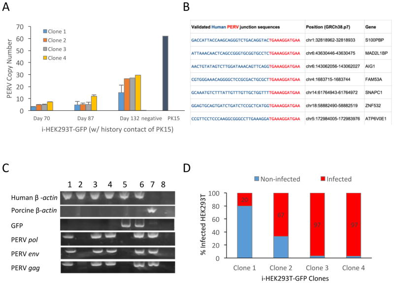
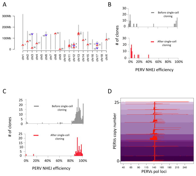
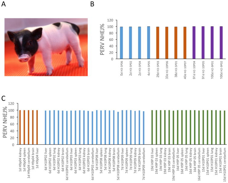
Xenotransplantation is a promising strategy to alleviate the shortage of organs for human transplantation. In addition to the concerns about pig-to-human immunological compatibility, the risk of cross-species transmission of porcine endogenous retroviruses (PERVs) has impeded the clinical application of this approach. We previously demonstrated the feasibility of inactivating PERV activity in an immortalized pig cell line. We now confirm that PERVs infect human cells, and we observe the horizontal transfer of PERVs among human cells. Using CRISPR-Cas9, we inactivated all of the PERVs in a porcine primary cell line and generated PERV-inactivated pigs via somatic cell nuclear transfer. Our study highlights the value of PERV inactivation to prevent cross-species viral transmission and demonstrates the successful production of PERV-inactivated animals to address the safety concern in clinical xenotransplantation.


Figures

Figure 1
Figure 1. Pig-to-human and human-to-human PERVs transmission
A) PERVs copy number in infected human cells increases over time when co-cultured with PK15 cells. Human HEK293T-GFP cells were co-cultured with equivalent numbers of pig PK15 cells for one week. HEK293T-GFP without any contact of PK15 cells were used as negative control (negative). B) Detection of PERVs insertion sites in human genome. Among the 22 PERV insertion sites detected by inverse PCR, 15 were mapped to the intragenic region. We tested a portion of the intragenic hits and validated 7 out of 12 by junction PCR (shown here). The 30bp human genomic sequences are shown in blue, whereas the PERV LTRs are shown in red. C) Detection of human-to-human PERVs transmission. Individual clones of HEK293T were grown from the single cells isolated from the co-culture of i-HEK293T-GFP with HEK293T through flow cytometry. The PCR gel image showed that 3 out of 4 randomly tested HEK293T clones were infected, which contained PERVs sequences (PERV pol, env, and gag), but no sequence of GFP or pig genomic DNA (tested by pig specific GGTA). Sample orders are: 1) HEK293T clone 1; 2) HEK293T clone 2; 3) HEK293T clone 3; 4) HEK293T clone 4; 5) HEK293T-GFP control; 6) i-HEK293T-GFP; 7) PK15 WT; and 8) negative. D) Four different i-HEK293T-GFP clones have different infectious potential. Four infected parental i-HEK293T-GFP clones are co-cultured with WT HEK293T. The PERV copy number of the 4 parental i-HEK293T-GFP clones are 15, 28, 27 and 28, respectively. The percentages of the infected WT HEK293T clones from the co-culture of i-HEK293T-GFP and WT HEK293T varied from 20% to 97%. Primers used are listed in the Methods Table 1.
Figure 2
Figure 2. PERVs insertion sites mapping, and genome-wide inactivation
A) Chromosome mapping of PERVs locations in FFF3. Chromosomal scaffolds are in grey. Red arrows represent PERVs in the forward or positive chain of chromosome. Blue arrows represent PERVs in the reverse or negative chain. Y-axis represents chromosomal coordinates. Two additional copies were mapped to repetitive regions, and two could not be mapped to the current pig genome assembly and are not shown (11% gaps, Sus scrofa build 10.2) (10). B) Failure to obtain 100% PERV-inactivated FFF3 clones using CRISPR-Cas9. After targeting the PERVs in FFF3, single cells were sorted and immediately genotyped. We observed a bimodal distribution of PERV targeting frequencies among single cells (upper panel), similar to that seen in the PK15 clones (5). 100% PERV-inactivated FFF3 cells were present among the single cells directly genotyped. However, this pattern changed after expansion of the single cells (bottom panel). Among the single cell clones, we only obtained the ones with lower efficiency (≤39%, the average targeting efficiency in the population was 37%), but not the ones with 100% PERV inactivation (lower panel). C) Treatment with PFTα and bFGF sustained the growth of highly modified FFF3 clones. The combined use of a p53 inhibitor, PFTα, and a growth factor, bFGF, rescued the highly modified cells. A population of FFF3 was treated with PFTα and bFGF during the gene editing experiment (Methods); then, single cells were sorted for direct genotyping and for colony growth followed by genotyping. Both the single cells and expanded clones showed similar distribution in PERV targeting efficiency, and highly modified clones survived under this condition. D) Genotype of 100% PERV inactivated clones. Several 100% PERV-inactivated clones were achieved from the PFTα and bFGF treated FFF3 population. The figure shows haplotypes of one of the 100% PERV-inactivated clones at PERV pol loci, after CRISPR-Cas9 treatment. The y-axis indicates the edited PERVs loci. The x-axis indicates the relative locations of the indels within the PERV loci. Aligned indel events in the PERV pol sequence are represented in red. Shades of purple indicate different haplotypes of PERVs.
Figure 3
Figure 3. PERV-inactivated pigs
A) Image of the first born PERV-inactivated pig. This picture showed the first born pig (Laika) at day 2 after birth. B) PERV inactivation at genomic DNA level. We genotyped PERV-inactivated pigs at different ages (up to 100 days) by deep sequencing of the PERV pol loci. All examined pigs showed ~100% PERV inactivation efficiency, which demonstrates that there is no detectable PERV reinfection from surrogate sows to cloned pigs. C) PERV inactivation at mRNA level. Total mRNA generated cDNA was used to detect the PERV inactivation efficiency of the PERV-ko pig transcripts. All pigs exhibited ~100% PERV eradication efficiency at mRNA level.

Comment in

References

    1. Shafran D, Kodish E, Tzakis A. Organ shortage: the greatest challenge facing transplant medicine. World J Surg. 2014;38:1650–7. - PubMed
    1. Deschamps JY, Roux FA, Saï P, Gouin E. History of xenotransplantation. Xenotransplantation. 2005;12:91–109. - PubMed
    1. Patience C, Takeuchi Y, Weiss RA. Infection of human cells by an endogenous retrovirus of pigs. Nat Med. 1997;3:282–6. - PubMed
    1. Denner J. How active are porcine endogenous retroviruses (PERVs)? Viruses. 2016;8:216. - PMC - PubMed
    1. Yang L, et al. Genome-wide inactivation of porcine endogenous retroviruses (PERVs) Science. 2015;350:1101–4. - PubMed

Publication types

MeSH terms