Review
. 2017 Aug 10;131(17):2257-2274.
doi: 10.1042/CS20160381. Print 2017 Sep 1.

Perivascular spaces, glymphatic dysfunction, and small vessel disease

Affiliations
Review

Perivascular spaces, glymphatic dysfunction, and small vessel disease

Humberto Mestre et al. Clin Sci (Lond). .

Abstract

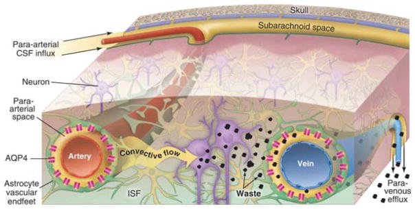
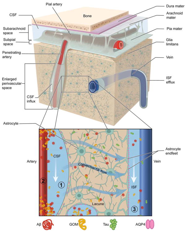
Cerebral small vessel diseases (SVDs) range broadly in etiology but share remarkably overlapping pathology. Features of SVD including enlarged perivascular spaces (EPVS) and formation of abluminal protein deposits cannot be completely explained by the putative pathophysiology. The recently discovered glymphatic system provides a new perspective to potentially address these gaps. This work provides a comprehensive review of the known factors that regulate glymphatic function and the disease mechanisms underlying glymphatic impairment emphasizing the role that aquaporin-4 (AQP4)-lined perivascular spaces (PVSs), cerebrovascular pulsatility, and metabolite clearance play in normal CNS physiology. This review also discusses the implications that glymphatic impairment may have on SVD inception and progression with the aim of exploring novel therapeutic targets and highlighting the key questions that remain to be answered.

Keywords: CNS clearance; Cerebrospinal fluid; Glymphatic; Perivascular space; Small vessel disease.


Conflict of interest statement

Conflicts of Interest

The authors declare no conflicts of interest.

Figures

Figure 1
Figure 1. Overview of the Glymphatic System
Convective flow of cerebrospinal fluid (CSF) enters the PVS of pial arteries and dives down into the brain through penetrating artery PVS. These PVS are surrounded on the outside by a sheath of astroglial endfeet lined with aquaporin-4 (AQP4) water channels. AQP4 facilitates the bulk flow movement of CSF into the parenchyma where it mixes with interstitial fluid (ISF). Convective ISF flow propels waste products, such as amyloid-β and tau, towards veins where they enter PVS for efflux out of the CNS. This process is regulated by changes in the extracellular space volume of the parenchyma as is seen during sleep-wake state transitions. Adapted with permission from the AAAS(126).
Figure 2
Figure 2. Potential Mechanisms Driving Glymphatic System Impairment in SVD
(1) PVS: Abluminal deposits of Aβ in CAA and GOM in CADASIL block PVS flows and may cause loss of AQP4 polarization and astrocyte endfoot and pericyte detachment. Increased PVS fluid and structural remodeling causes enlarged PVS. (2) Cerebrovascular pulsatility: Vascular pathology causes a decrease in vessel wall compliance increasing cerebrovascular pulsatility and mechanical stress against the PVS. Diffuse endothelial cell failure and impaired neurovascular unit cross-talk results in a net decrease in ISF formation offering a larger driving force for CSF entry. (3) CNS Clearance: Parenchymal changes associated to injury such as astrogliosis and lacunar infarcts, change the extracellular space volume and tortuosity, predisposing to solute trapping and reduced clearance of toxic metabolic byproducts (e.g. Aβ and tau). These changes would also decrease ISF efflux and cause fluid stasis. PVS inflammation due to extravasation of plasma proteins gives rise to perivascular edema and WML.

References

    1. Hachinski V World Stroke O. Stroke and Potentially Preventable Dementias Proclamation: Updated World Stroke Day Proclamation. Stroke. 2015;46(11):3039–40. - PubMed
    1. Wardlaw JM, Smith EE, Biessels GJ, Cordonnier C, Fazekas F, Frayne R, et al. Neuroimaging standards for research into small vessel disease and its contribution to ageing and neurodegeneration. Lancet Neurol. 2013;12(8):822–38. - PMC - PubMed
    1. Wardlaw JM, Smith C, Dichgans M. Mechanisms of sporadic cerebral small vessel disease: insights from neuroimaging. Lancet Neurol. 2013;12(5):483–97. - PMC - PubMed
    1. Charidimou A, Pantoni L, Love S. The concept of sporadic cerebral small vessel disease: A road map on key definitions and current concepts. Int J Stroke. 2016;11(1):6–18. - PubMed
    1. Pantoni L. Cerebral small vessel disease: from pathogenesis and clinical characteristics to therapeutic challenges. Lancet Neurol. 2010;9(7):689–701. - PubMed

LinkOut - more resources