. 2017 Oct 6;292(40):16665-16676.
doi: 10.1074/jbc.M117.803320. Epub 2017 Aug 10.

Conformational characterization of nerve growth factor-β reveals that its regulatory pro-part domain stabilizes three loop regions in its mature part

Affiliations

Conformational characterization of nerve growth factor-β reveals that its regulatory pro-part domain stabilizes three loop regions in its mature part

Esben Trabjerg et al. J Biol Chem. .

Abstract

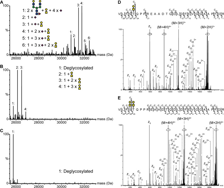
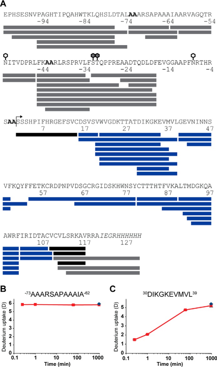
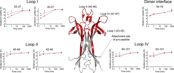
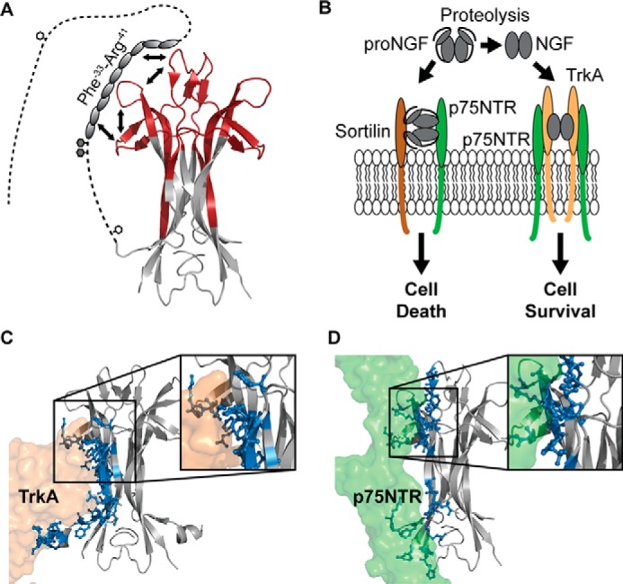
Nerve growth factor-β (NGF) is essential for the correct development of the nervous system. NGF exists in both a mature form and a pro-form (proNGF). The two forms have opposing effects on neurons: NGF induces proliferation, whereas proNGF induces apoptosis via binding to a receptor complex of the common neurotrophin receptor (p75NTR) and sortilin. The overexpression of both proNGF and sortilin has been associated with several neurodegenerative diseases. Insights into the conformational differences between proNGF and NGF are central to a better understanding of the opposing mechanisms of action of NGF and proNGF on neurons. However, whereas the structure of NGF has been determined by X-ray crystallography, the structural details for proNGF remain elusive. Here, using a sensitive MS-based analytical method to measure the hydrogen/deuterium exchange of proteins in solution, we analyzed the conformational properties of proNGF and NGF. We detected the presence of a localized higher-order structure motif in the pro-part of proNGF. Furthermore, by comparing the hydrogen/deuterium exchange in the mature part of NGF and proNGF, we found that the presence of the pro-part in proNGF causes a structural stabilization of three loop regions in the mature part, possibly through a direct molecular interaction. Moreover, using tandem MS analyses, we identified two N-linked and two O-linked glycosylations in the pro-part of proNGF. These results advance our knowledge of the conformational properties of proNGF and NGF and help provide a rationale for the diverse biological effects of NGF and proNGF at the molecular level.

Keywords: glycoprotein; hydrogen exchange mass spectrometry; hydrogen/deuterium exchange; intrinsically disordered protein; neurodegeneration; neurodegenerative disease; neurotrophin; protein conformation; protein structure.


Conflict of interest statement

The authors declare that they have no conflicts of interest with the contents of this article

Figures

Figure 1.
Figure 1.
Structure of NGF. A, crystal structure of the NGF dimer (PDB ID: 1SG1). The cysteine knot is highlighted in yellow. B, schematic representation of the translation product of the NGF gene. Residues in the signal and the pro-part of proNGF are by convention assigned negative numbers (21). The connectivity of the three cysteine bonds in the mature part is shown. White bar, signal peptide of NGF; light gray bar, pro-part of NGF; dark gray bar, mature NGF.
Figure 2.
Figure 2.
Identification and mapping of N- and O-linked glycans in the pro-part of proNGF. Intact mass analysis of nontreated (A), PNGase- and sialidase-treated (B), and PNGase F-, sialidase-, and O-glycosidase-treated (C) proNGF. Treatment with both PNGase F and O-glycosidase caused a mass shift corresponding to known glycan structures, showing that proNGF is both N- and O-linked glycosylated. The ETD fragmentation spectra of glycopeptide Val−35–Arg−7 with either two (E) or one (D) O-linked glycan show that proNGF is O-linked glycosylated at Ser−32 and Thr−31. The mass error of all identified peaks is below 23 ppm. Yellow square, N-acetylgalactosamine; blue square, N-acetylglucosamine; yellow circle, galactose; green circle, mannose; purple diamond, sialic acid; red triangle, fucose.
Figure 3.
Figure 3.
HDX-MS coverage map of proNGF and NGF. A, peptic peptides from which HDX data could be obtained are shown as: blue bars, data for both proNGF and NGF; gray bars, data only for proNGF; and black bars, data for NGF only. The bent arrow marks position 0, i.e. the beginning of the mature part of NGF. Glycosylations in the pro-part of proNGF are marked by white (N-linked) and gray (O-linked) hexagons. The alanine mutations at positions −73, −72, −43, −42, −2, and −1 are highlighted by a bold font, and the His6 tag is highlighted by an italic font. The graphs show absolute deuterium incorporation, plotted as a function of time, of a disordered region from the pro-part (B) and a structured region in the mature part (C) of proNGF. Blue-filled circles, deuterium incorporation of equilibrium labeled (90%) samples. S.D. is plotted as error bars (only slightly visible) (n = 3 for the 15-s, 1-min, and 60-min time points, and n = 2 for the 16-h time point and the equilibrated control sample).
Figure 4.
Figure 4.
Detection of local higher-order structure in the pro-part of proNGF. A, deuterium incorporation after 15 s (red bars) was compared with deuterium incorporation of equilibrium labeled (90%) samples (blue bars). All peptides shown originated from the pro-part of proNGF. A significantly lesser incorporation of deuterium was seen from residue Arg−41–Phe−33. Glycopeptides was grouped by themselves, as a possible protection could be assigned to both the peptide backbone and the attached glycans (72). Asterisks, indicate significant differences in exchange (p < 0.01). S.D. is plotted as error bars. a, peptide contains one O-linked glycan; b, peptide contains two O-linked glycans. (n = 3 for the 15-s time point, and n = 2 for the equilibrated control sample). B, structure prediction of the pro-part of proNGF performed by several algorithms. DISOPRED2 predicts whether a residue is natively disordered or structured, whereas PSIPRED v3.3 and JPRED 3 predict the presence of secondary structure elements, e.g. random coil, α-helix, and β-strand. The lack of disorder predicted for residues Arg−47–Ser−32 aligns well with the observed protection from exchange in the region of residues Arg−41–Phe−33. Gray bars, predicted disordered regions; green bars, predicted α-helix structure; yellow bars, predicted β-sheet structure. AA, double alanine mutation site; white hexagon, N-linked glycosylation site; gray hexagon, O-linked glycosylation site.
Figure 5.
Figure 5.
The pro-part of proNGF perturbs the conformation of the mature part of proNGF. Absolute deuterium incorporation is plotted as a function of time for NGF (gray lines) and the mature part of proNGF (red lines). Equilibrium-labeled (90%) proNGF (control samples) are plotted as blue-filled circles at the 16-h time point. Regions in the mature part of proNGF where a significant protection of exchange was observed in the presence of the pro-part for at least two consecutive time points are colored red on a crystal structure of NGF (PDB ID: 1SG1). S.D. is plotted as error bars (most are too small to be visible). Asterisk, denotes a significant protection from exchange when comparing deuterium incorporation in NGF with the mature part of proNGF (p < 0.01). (n = 3 for the 15-s, 1-min, and 60-min time points, and n = 2 for the 16-h time point and the equilibrated control sample).
Figure 6.
Figure 6.
A refined structural model of proNGF. A, schematic representation of the findings presented in the current study. We propose that the part of the pro-part where local higher-order structure was identified interacts with parts of loops I, II, and IV via a combined intra- and intermolecular mechanism. The white hexagons mark N-linked glycosylation sites, and the gray hexagons mark O-linked glycosylation sites. To decrease the complexity, the pro-part was included on only one of the NGF monomers (PDB ID: 1SG1). B, biological effect of proNGF and NGF on neurons. NGF induces the growth of neurons by binding to the TrkA and the p75NTR receptors, whereas proNGF mediates apoptosis of neurons by binding to a receptor complex of sortilin and p75NTR. Figure adapted from (8). C and D, crystal structures of NGF (gray) in complex with TrkA (orange) and p75NTR (green) (PDB ID: 2IFG and 1SG1). Residues in NGF that form hydrogen bonds with the receptors are highlighted as sticks and colored blue. Inserts, zoom image of the binding surface implicating loops I, II, and IV of NGF. To simplify this figure, the second receptor molecule of TrkA and p75NTR was omitted.

References

    1. Chao M. V. (2003) Neurotrophins and their receptors: A convergence point for many signalling pathways. Nat. Rev. Neurosci. 4, 299–309 - PubMed
    1. Jansen P., Giehl K., Nyengaard J. R., Teng K., Lioubinski O., Sjoegaard S. S., Breiderhoff T., Gotthardt M., Lin F., Eilers A., Petersen C. M., Lewin G. R., Hempstead B. L., Willnow T. E., and Nykjaer A. (2007) Roles for the pro-neurotrophin receptor sortilin in neuronal development, aging and brain injury. Nat. Neurosci. 10, 1449–1457 - PubMed
    1. Nykjaer A., Willnow T. E., and Petersen C. M. (2005) p75NTR: Live or let die. Curr. Opin. Neurobiol. 15, 49–57 - PubMed
    1. Seidah N. G., Benjannet S., Pareek S., Savaria D., Hamelin J., Goulet B., Laliberte J., Lazure C., Chrétien M., and Murphy R. A. (1996) Cellular processing of the nerve growth factor precursor by the mammalian pro-protein convertases. Biochem. J. 314, 951–960 - PMC - PubMed
    1. Paoletti F., Malerba F., Kelly G., Noinville S., Lamba D., Cattaneo A., and Pastore A. (2011) Conformational plasticity of proNGF. PLoS ONE 6, e22615. - PMC - PubMed

Publication types

LinkOut - more resources