

A rare urological presentation of appendicitis
- PMID: 28798242
- PMCID: PMC5747616
- DOI: 10.1136/bcr-2017-220546
A rare urological presentation of appendicitis
Abstract
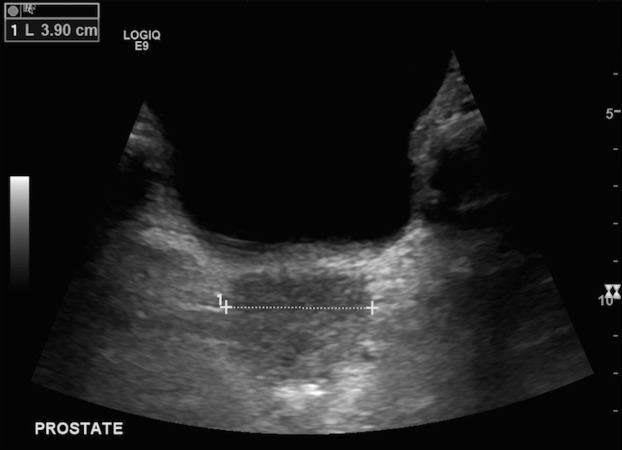
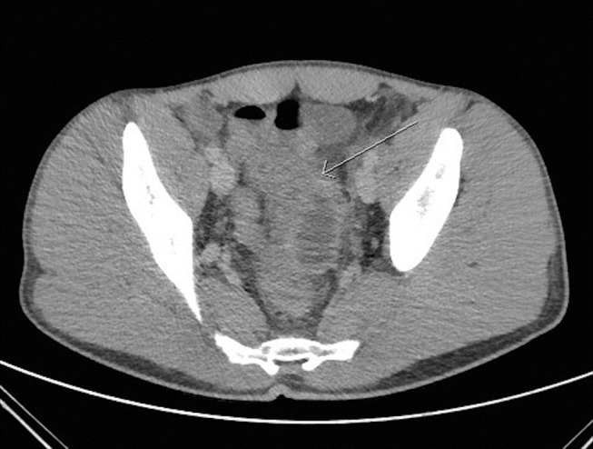
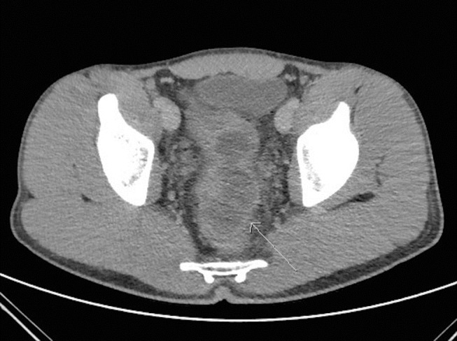
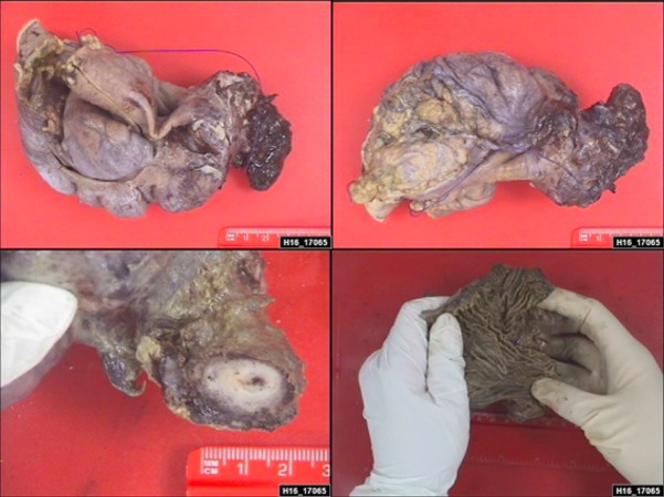
A 17-year-old boy with no medical comorbidities, but a significant family history of malignancy, presented to Accident and Emergency following 3 days of increasing rectal pain, symptoms of bladder outflow obstruction (poor flow, intermittent stream and hesitancy) and dysuria. Notably he had no abdominal pain. Digital rectal examination revealed a tender, enlarged prostate. Inflammatory markers were significantly raised (white cell count 17.7, C reactive protein 191). He was diagnosed clinically as prostatitis and commenced on intravenous antibiotics. Despite this his pain and inflammatory markers deteriorated, necessitating a CT of his abdomen and pelvis. This demonstrated multiloculated large thick-walled abscesses in the pelvis closely related to the rectum, prostate and seminal vesicles with some bowel wall thickening. Laparoscopy demonstrated a large colonic mass adherent to surrounding structures. The procedure was converted to laparotomy to enable resection of the mass via a limited right haemicolectomy. He recovered well and was discharged. Histopathological analysis of the specimen revealed appendicitis.
Keywords: gastrointestinal Surgery; general surgery; prostate; urinary and genital tract disorders.
© BMJ Publishing Group Ltd (unless otherwise stated in the text of the article) 2017. All rights reserved. No commercial use is permitted unless otherwise expressly granted.
Conflict of interest statement
Competing interests: None declared.
Figures
References
-
- Jones WG, Barie PS. Urological manifestations of acute appendicitis. J Urol 1988;139:1325–8. doi:10.1016/S0022-5347(17)42911-9 - DOI - PubMed
-
- Guidry SP, Poole GV. The anatomy of appendicitis. Am Surg 1994;60:68–71. - PubMed
-
- Tundidor Bermúdez AM, Amado Diéguez JA. Montes De Oca Mastrapa jl.urological manifestations of acute appendicitis. Arch Esp Urol 2005;58:207–12. - PubMed
-
- Humes DJ, Simpson J. Acute appendicitis. BMJ 2006;333:530–4. doi:10.1136/bmj.38940.664363.AE - DOI - PMC - PubMed
Publication types
MeSH terms
LinkOut - more resources
Full Text Sources
Other Literature Sources
Medical
Research Materials