. 2017 Aug 10;7(1):7831.
doi: 10.1038/s41598-017-08314-1.

Sphingomyelin as a myelin biomarker in CSF of acquired demyelinating neuropathies

Affiliations

Sphingomyelin as a myelin biomarker in CSF of acquired demyelinating neuropathies

Giovanna Capodivento et al. Sci Rep. .

Abstract

Fast, accurate and reliable methods to quantify the amount of myelin still lack, both in humans and experimental models. The overall objective of the present study was to demonstrate that sphingomyelin (SM) in the cerebrospinal fluid (CSF) of patients affected by demyelinating neuropathies is a myelin biomarker. We found that SM levels mirror both peripheral myelination during development and small myelin rearrangements in experimental models. As in acquired demyelinating peripheral neuropathies myelin breakdown occurs, SM amount in the CSF of these patients might detect the myelin loss. Indeed, quantification of SM in 262 neurological patients showed a significant increase in patients with peripheral demyelination (p = 3.81 * 10 - 8) compared to subjects affected by non-demyelinating disorders. Interestingly, SM alone was able to distinguish demyelinating from axonal neuropathies and differs from the principal CSF indexes, confirming the novelty of this potential CSF index. In conclusion, SM is a specific and sensitive biomarker to monitor myelin pathology in the CSF of peripheral neuropathies. Most importantly, SM assay is simple, fast, inexpensive, and promising to be used in clinical practice and drug development.


Conflict of interest statement

The authors declare that they have no competing interests.

Figures
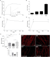
Figure 1
Figure 1
SM levels mirror both peripheral myelination during development and small myelin rearrangements (A) SM titration curve of a fluorescence-based assay showed a significant and reliable linearity from 0.2 to 1.6 nmol. (B) Quantification of SM in several tissues from 270-day-old rats displayed a specific enrichment in sciatic nerves. (C,D) SM levels in rat sciatic nerves and purified myelin progressively increased during development (sciatic nerve: 3-day-old, n = 5; 30-day-old, n = 8; 365-day-old, n = 3; purified myelin: 3-day-old, n = 3; 40-day-old, n = 3; 300-day-old, n = 3). (E). We treated DRG cultures with Forskolin (Fsk) 20 and 40 μM to induce demyelination and then let them recover by Fsk removal. We found that SM was able to detect both demyelination and remyelination (n = 6). (F,G) Immunofluorescence staining of myelin by MBP in DRG cultures and quantification of MBP-positive myelinated fibers supported biochemical data. Scale bars: 100 µm. Errors bars in A, B represent the SD; *p < 0.05, **p < 0.01, ***p < 0.001, ****p < 0.0001, ns = not-significant. Goodness of fit (r2) was determined by linear regression analysis. Holm-Sidak multiple comparison test after 1-way analysis of variance was used for statistical comparison in B-E.
Figure 2
Figure 2
SM is increased in patients affected by demyelinating neuropathies. (A,B) We assessed SM in clinically well-characterized subgroups of patients affected by: definite demyelinating neuropathies, namely GBS and CIDP (Black bar, Demyelinating neuropathies, n = 14), neurological disorders with blood brain barrier dysfunction but not overt demyelination (Grey bar, BBB dysfunction, n = 13), and other non-demyelinating neurological disorders (White bar, Ctrl, n = 15). We demonstrated that SM was significantly increased in patients with demyelinating neuropathies compared to both the control groups. Interestingly, SM exclusively increased in the CSF of these patients to exclude a direct exchange with peripheral blood. (C,D) LC-MS/MS targeted-sphingomyelin analysis showed that the total SM species contribution is definitely consistent with those obtained by the fluorescence-based assay. 16:0, 18:0, 24:0, 24:1 are the most abundant SM species with different saturated and unsaturated fatty acid chains. (E) Specificity and sensitivity of SM assay were tested by ROC curve analysis (F) A cut-off value for SM was also calculated. ***p < 0.001, ****p < 0.0001, ns = not-significant. Holm-Sidak multiple comparison test after 1-way analysis of variance was used for statistical comparison in (AD). ROC implementation in Mathworks MATLAB environment was used in E, F.
Figure 3
Figure 3
SM is able to distinguish axonal from demyelinating neuropathies. SM alone was significantly increased in patients affected by demyelinating neuropathies (n = 14) compared to those affected by axonal neuropathies (n = 8). ***p < 0.001. Unpaired 2-tailed t-test was used for statistical comparison.
Figure 4
Figure 4
SM is a biomarker of myelin breakdown in the CSF of human patients. A scatter plot of all the 262 analysed patients based on QAlb and SM cut-off (X and Y axis, respectively) is shown. Notably, most of the patients with peripheral demyelination (Black circles) were over the SM threshold. Control subjects, either with or without BBB dysfunction (Grey and white circles, respectively) were also reported in the graph. Grey lines represent SM (Y axis) and QAlb (X axis) cut-off.

References

    1. Najm FJ, et al. Drug-based modulation of endogenous stem cells promotes functional remyelination in vivo. Nature. 2015;522:216–220. doi: 10.1038/nature14335. - DOI - PMC - PubMed
    1. Lesko LJ, Atkinson AJ., Jr. Use of biomarkers and surrogate endpoints in drug development and regulatory decision making: criteria, validation, strategies. Annu Rev Pharmacol Toxicol. 2001;41:347–366. doi: 10.1146/annurev.pharmtox.41.1.347. - DOI - PubMed
    1. Wang Y, et al. Differentiation and quantification of inflammation, demyelination and axon injury or loss in multiple sclerosis. Brain. 2015;138:1223–1238. doi: 10.1093/brain/awv046. - DOI - PMC - PubMed
    1. Bodini B, Veronese M, Turkheimer F, Stankoff B. Benzothiazole and stilbene derivatives as promising positron emission tomography myelin radiotracers for multiple sclerosis. Ann Neurol. 2016;80:166–167. doi: 10.1002/ana.24667. - DOI - PubMed
    1. Dortch RD, Dethrage LM, Gore JC, Smith SA, Li J. Proximal nerve magnetization transfer MRI relates to disability in Charcot-Marie-Tooth diseases. Neurology. 2014;83:1545–1553. doi: 10.1212/WNL.0000000000000919. - DOI - PMC - PubMed

Publication types

MeSH terms