. 2017 Aug 10;7(1):7797.
doi: 10.1038/s41598-017-07513-0.

MicroRNA-132 promotes fibroblast migration via regulating RAS p21 protein activator 1 in skin wound healing

Affiliations

MicroRNA-132 promotes fibroblast migration via regulating RAS p21 protein activator 1 in skin wound healing

Xi Li et al. Sci Rep. .

Abstract

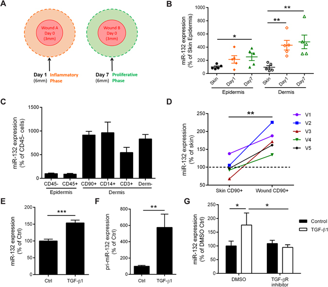
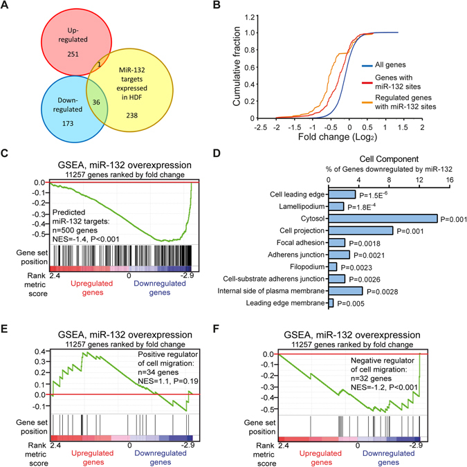
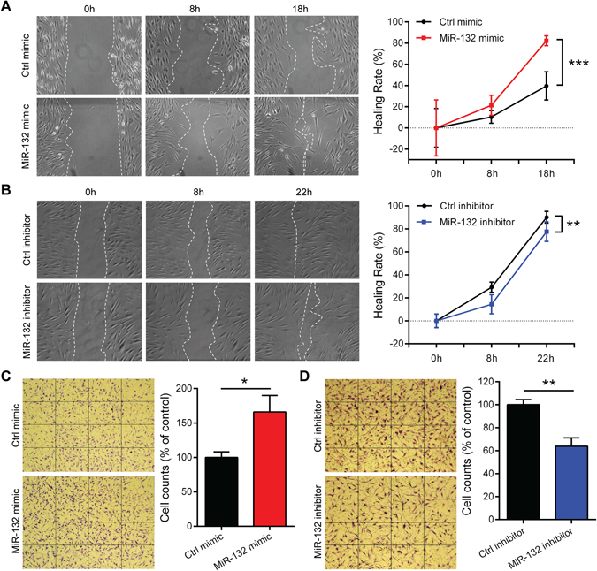
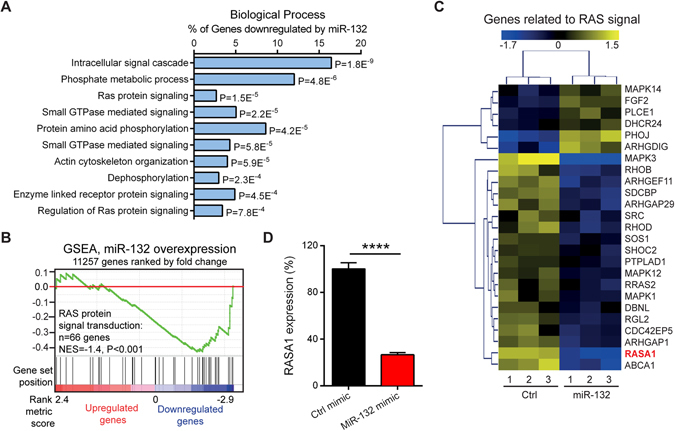
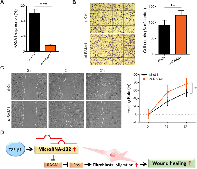
MicroRNA (miR)-132 has been identified as a top up-regulated miRNA during skin wound healing and its inhibition impairs wound repair. In a human in vivo surgical wound model, we showed that miR-132 was induced in epidermal as well as in dermal wound-edge compartments during healing. Moreover, in a panel of cells isolated from human skin wounds, miR-132 was found highly expressed in human dermal fibroblasts (HDFs). In HDFs, miR-132 expression was upregulated by TGF-β1. By overexpression or inhibition of miR-132, we showed that miR-132 promoted HDF migration. Mechanistically, global transcriptome analysis revealed that RAS signaling pathway was regulated by miR-132 in HDFs. We found that RAS p21 protein activator 1 (RASA1), a known target of miR-132, was downregulated in HDFs upon miR-132 overexpression. Silencing of RASA1 phenocopied the pro-migratory effect of miR-132. Collectively, our study reveals an important role for miR-132 in HDFs during wound healing and indicates a therapeutic potential of miR-132 in hard-to-heal skin wounds.


Conflict of interest statement

The authors declare that they have no competing interests.

Figures

Figure 1
Figure 1
Expression and regulation of miR-132 in wounds. (A) The intact skin (Day 0), Day 1 and Day 7 wounds were collected from healthy donors. (B) MiR-132 expression was analyzed in the epidermis and dermis of the biopsies from donor 1–5, which were separated by laser capture microdissection. (C) QRT-PCR analysis of miR-132 expression in the cells isolated from the Day6 wounds of 5 donors, including CD45- and CD45 + cells from epidermis, and CD90 + , CD14 + , CD3 + and CD90-CD14-CD3- (Derm-) cells from dermis. (D) QRT-PCR analysis of miR-132 expression in dermal CD90 + cells from intact skin and Day6 wounds. V1–5 indicate 5 healthy volunteers. QRT-PCR analysis of miR-132 (E) or its primary precursor pri-miR-132 (F) in HDFs treated with 10 nM TGF-β1 for 48 hours (n = 3). (G) The TGF-β receptor inhibitor SB431542 was applied 15 minutes before adding TGF-β1 to HDFs, and miR-132 expression was analyzed 48 hours later (n = 3). The data are presented as mean ± s.e.m. in (B, C), as mean ± s.d. in (E-G). *P < 0.05, **P < 0.01 and ***P < 0.001 by Student’s t test.
Figure 2
Figure 2
Transcriptome analysis of fibroblasts overexpressing miR-132. (A) Venn diagram depicting the number of genes significantly up- (red) or down-regulated (blue) by miR-132 (fold change ≥ 1.5, P <0.05) and TargetScan predicted miR-132 targets that expressed in HDFs (yellow). (B) Cumulative distribution plots of log2-transformed fold-changes for genes predicted to contain miR-132 binding sites (red), genes with miR-132 sites and regulated by miR-132 (orange) and all the genes detected by microarray (blue). (C) Genes in microarray data were ranked by fold change (miR-132 mimic / Ctrl mimic). GSEA evaluated enrichment within the profile data for the predicted target genes of miR-132. Vertical bars along the χ axis denote the positions of miR-132 target genes within the ranked list. NES, normalized enrichment score. (D) The top10 gene ontology cell component terms for the genes down-regulated by miR-132 in fibroblasts. P-values were determined by the Fisher’s exact test. GSEA evaluated enrichment within the microarray data for the genes reported to accelerate (E) or impair (F) cell migration.
Figure 3
Figure 3
MiR-132 promotes fibroblast migration. Scratch assays were performed to assess the migration rate of HDFs transfected with 20 nM miR-132 mimics (A) or miR-132 inhibitors (B) for 48 hours. Photographs were taken at indicated time points after scratch injury. The healing rates were quantified by measuring the area of the injured region. **P < 0.01 and***P < 0.001 by Repeated Measures Two-way ANOVA. Transwell migration assay were performed to assess the motility of fibroblasts transfected with 20 nM miR-132 mimics (C) or miR-132 inhibitors (D). The number of fibroblasts passing through the membrane within 24 hours was counted. *P < 0.05 and **P < 0.01 by Student’s t-test. The data are presented as mean ± s.d.
Figure 4
Figure 4
MiR-132 regulates RAS signalling pathway. (A) The top10 gene ontology biological process terms for the genes down-regulated by miR-132 in fibroblasts. P-values were determined by the Fisher’s exact test. (B) GSEA evaluated enrichment for the genes related to the RAS signalling within the microarray data. (C) The genes related to the RAS signalling were regulated by miR-132 in fibroblasts. Heat map illustrates levels of significantly changed genes (fold change ≥ 1.2, P < 0.05). Colour intensity is scaled within each row so that the highest expression value corresponds to bright yellow and the lowest to bright blue. (D) RASA1 was detected by qRT-PCR in HDFs transfected with 20 nM miR-132 mimics or control oligos (n = 3). The data are presented as mean ± s.d. ****P < 0.0001 by Student’s t test.
Figure 5
Figure 5
MiR-132 promotes migration by regulating RASA1. (A) RASA1 knockdown efficiency was analysed by qRT-PCR in HDFs transfected with 20 nM RASA1 specific siRNAs or control siRNAs (n = 3). (B) Transwell migration assay was performed in HDFs with silenced RASA1 expression (n = 3). **P < 0.01 and ***P < 0.001 by Student’s t test. (C) Scratch assay was performed in HDFs with silenced RASA1 expression. *P < 0.05 by Repeated Measures Two-way ANOVA. The data are presented as mean ± s.d. (D) Schematic summary of the role of miR-132 in dermal fibroblasts during skin wound healing.

References

    1. Reinke JM, Sorg H. Wound repair and regeneration. Eur Surg Res. 2012;49:35–43. doi: 10.1159/000339613. - DOI - PubMed
    1. Gottrup F. A specialized wound-healing center concept: importance of a multidisciplinary department structure and surgical treatment facilities in the treatment of chronic wounds. Am J Surg. 2004;187:38S–43S. doi: 10.1016/S0002-9610(03)00303-9. - DOI - PubMed
    1. Frykberg RG, Banks J. Challenges in the Treatment of Chronic Wounds. Adv Wound Care (New Rochelle) 2015;4:560–582. doi: 10.1089/wound.2015.0635. - DOI - PMC - PubMed
    1. Gottrup F, Holstein P, Jorgensen B, Lohmann M, Karlsmar T. A new concept of a multidisciplinary wound healing center and a national expert function of wound healing. Arch Surg. 2001;136:765–772. doi: 10.1001/archsurg.136.7.765. - DOI - PubMed
    1. Richmond NA, Maderal AD, Vivas AC. Evidence-based management of common chronic lower extremity ulcers. Dermatol Ther. 2013;26:187–196. doi: 10.1111/dth.12051. - DOI - PubMed

Publication types

LinkOut - more resources