. 2017 Aug 10;7(1):7814.
doi: 10.1038/s41598-017-08412-0.

3D biomimetic artificial bone scaffolds with dual-cytokines spatiotemporal delivery for large weight-bearing bone defect repair

Affiliations

3D biomimetic artificial bone scaffolds with dual-cytokines spatiotemporal delivery for large weight-bearing bone defect repair

Xiaogang Bao et al. Sci Rep. .

Abstract

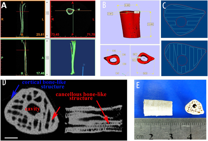
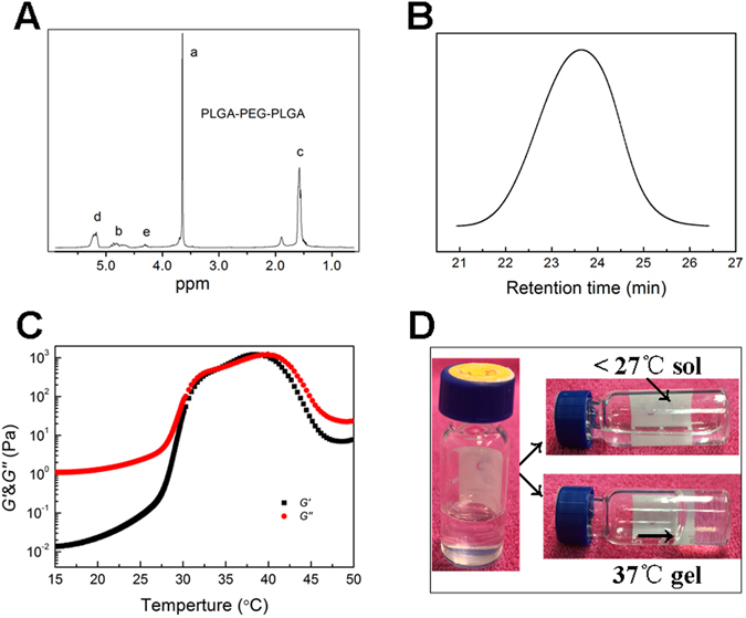
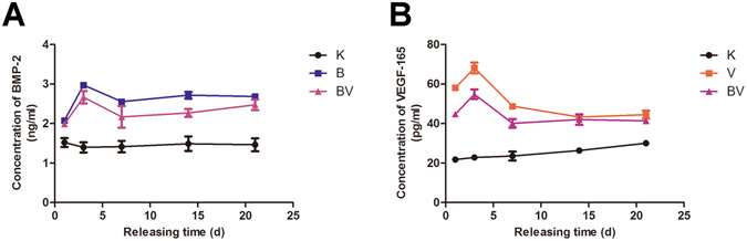
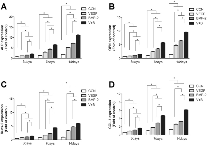
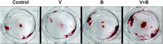
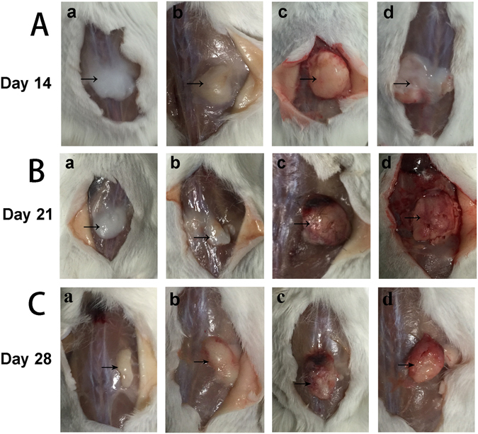
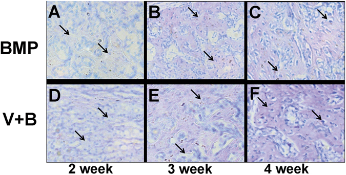
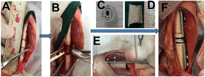
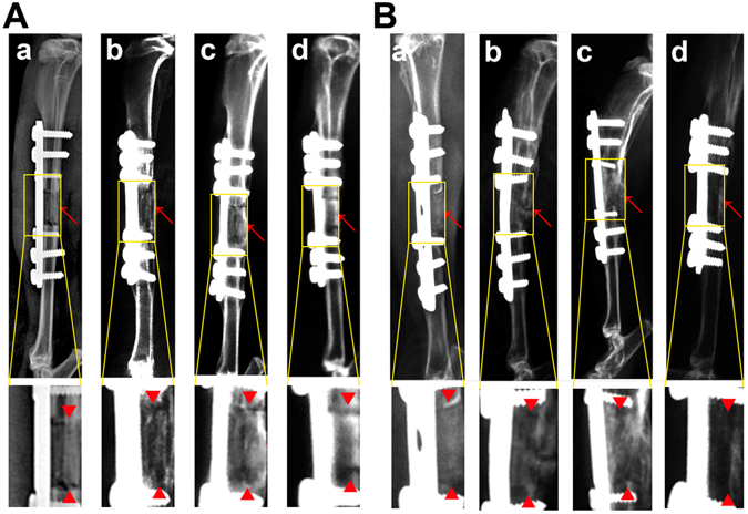
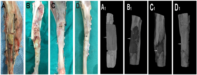
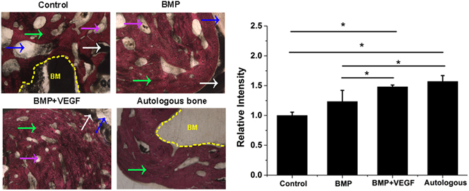
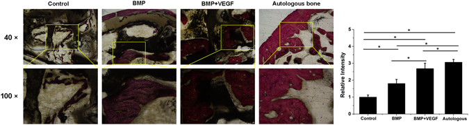
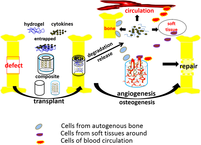
It is a great challenge to prepare "functional artificial bone" for the repair of large segmental defect, especially in weight-bearing bones. In this study, bioactive HA/PCL composite scaffolds that possess anatomical structure as autogenous bone were fabricated by CT-guided fused deposition modeling technique. The scaffolds can provide mechanical support and possess osteoconduction property. Then the VEGF-165/BMP-2 loaded hydrogel was filled into biomimetic artificial bone spatially to introduce osteoinduction and angioinduction ability via sustained release of these cytokines. It has been revealed that the cytokine-loaded hydrogel possessed good biodegradability and could release the VEGF-165/BMP-2 sustainedly and steadily. The synergistic effect of these two cytokines showed significant stimulation on the osteogenic gene expresssion of osteoblast in vitro and ectopic ossification in vivo. The scaffolds were then implanted into the rabbit tibial defect sites (1.2 cm) for bone regeneration for 12 weeks, indicating the best repair of defect in vivo, which was superior to the pure hydrogel/scaffolds or one-cytokine loaded hydrogel/scaffolds and close to autogenous bone graft. The strategy to construct an "anatomy-structure-function" trinity system as functional artificial bone shows great potential in replacing autogenous bone graft and applying in large bone defect repair clinically in future.


Conflict of interest statement

The authors declare that they have no competing interests.

Figures

Figure 1
Figure 1
(A) CT scanning of rabbit leg, (B) reconstruction of 1.2 cm-long tibia structure, (C) the fabrication path of FDM machine, (D) CT scanning of prepared artificial bone scaffolds, (E) The photograph of artificial bone scaffolds.
Figure 2
Figure 2
(A) 1H NMR specturm of the PLGA-PEG-PLGA copolymer, (B). GPC analysis of the copolymer shows a unimodal distribution, (C) the sol-gel transition process of the copolymer-saline solution (transition temperature of 35 °C), (D) the apperance of copolymer-saline solution at different temperatures.
Figure 3
Figure 3
(A) BMP-2 release in PBS of different groups for 21 days, (B). VEGF-165 release in PBS of different groups for 21 days. In the figure K represents control group- pure hydrogel, B for BMP-2 loaded-hydrogel, V for VEGF-165 loaded-hydrogel and BV for hydrogel loaded with both BMP-2 and VEGF-165.
Figure 4
Figure 4
The ALP (A), COL I (B), Runx-2 (C) and OPN (D) gene expression of MC3T3 cells cultured on hydrogels of control, V, B and VB groups.
Figure 5
Figure 5
The formation of calcium nodule of the four groups by Alizarin Red S Staining.
Figure 6
Figure 6
Hydrogel degradation under subcutaneous layer for 2 weeks (Line A), 3 weeks (Line B) and 4 weeks (Line C). lowercase letter a, b, c, d represent control group, V, B and VB group, respectively. Formation of bone-like hard tissue can be observed in B and VB group.
Figure 7
Figure 7
Toluidine blue staining of bone-like tissue B and BV group further proved the bone-like tissue formation.(AC) are analysis after injection of hydrogel for 2, 3 and 4 weeks for (B) group and (DF) are that of 2, 3 and 4 weeks for BV group, respectively.
Figure 8
Figure 8
The operation process of aritificial bone scaffold in rabbit tibial defect. (A) exposure of surgical field, (B) construction of bone defect (1.2 cm), (C)anatomical biomimetic artificial bone scaffold, (D) osteotomy of autogenous bone, (E) fixation of miniplate and screw fixation system, (F) successful bone graft transplant into bone defect.
Figure 9
Figure 9
X-ray scanning of control (a), B (b), VB (c) groups and autogenous bone (d) after implantation for 4 weeks (A) and 12 weeks (B). The lower images were magnified from the corresponding regions. Red arrows indicate the bone defect edges.
Figure 10
Figure 10
Gross appearance and CT reconstruction images of control (A,A 1), B (B,B 1), BV (C,C 1) groups and autogenous bone (D,D 1) for 12 weeks, arrows indicate the bone graft.
Figure 11
Figure 11
The VG staining of newly formed bone on the interface of implanted scaffolds/autogenous bone and nature bone at defect site of four groups (40×) with relevant statistics analysis. Pink arrow refers to PCL pieces, White arrow refers to HA pieces, Blue arrow refers to artificial scaffold,green arrow refers to new bone (NB), yellow area refers to bone marrow cavity (BM).
Figure 12
Figure 12
The VG staining of newly formed bone in the middle part inside the implanted scaffolds/autogenous bone of the four groups with relevant statistics analysis. The lower images were magnified from the corresponding regions.
Figure 13
Figure 13
Cytokines-loaded hydrogel was combined with the CT-guided 3D-printing artificial bone scaffolds and implanted into the complete tibial defect (1.2 cm). With the mechanical support and biocompatibility of the porous scaffolds, the sustained release of cytokines and the stimulation on angiogenesis and osteogenesis, the defect was successfully repaired.

References

    1. O’Keefe RJ, Mao J. Bone tissue engineering and regeneration: from discovery to the clinic–an overview. Tissue Eng Part B Rev. 2011;17:389–92. doi: 10.1089/ten.teb.2011.0475. - DOI - PMC - PubMed
    1. Soucacos PN, Dailiana Z, Beris AE, Johnson EO. Vascularised bone grafts for the management of non-union. Injury. 2006;37(Suppl 1):S41–50. doi: 10.1016/j.injury.2006.02.040. - DOI - PubMed
    1. Liu Y, Lim J, Teoh SH. Review: development of clinically relevant scaffolds for vascularised bone tissue engineering. Biotechnol Adv. 2013;31:688–705. doi: 10.1016/j.biotechadv.2012.10.003. - DOI - PubMed
    1. Fillingham Y, Jacobs J. Bone grafts and their substitutes. Bone Joint J. 2016;98-B:6–9. doi: 10.1302/0301-620X.98B.36350. - DOI - PubMed
    1. Einhorn TA, Gerstenfeld LC. Fracture healing: mechanisms and interventions. Nature Reviews. Rheumatology. 2015;11:45–54. - PMC - PubMed

Publication types

Substances

LinkOut - more resources