. 2017 Aug 10;7(1):7735.
doi: 10.1038/s41598-017-05245-9.

HIV-1 Tat-induced diarrhea evokes an enteric glia-dependent neuroinflammatory response in the central nervous system

Affiliations

HIV-1 Tat-induced diarrhea evokes an enteric glia-dependent neuroinflammatory response in the central nervous system

Giuseppe Esposito et al. Sci Rep. .

Abstract

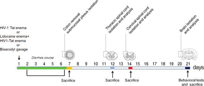
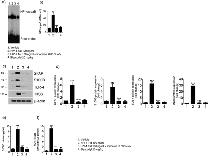
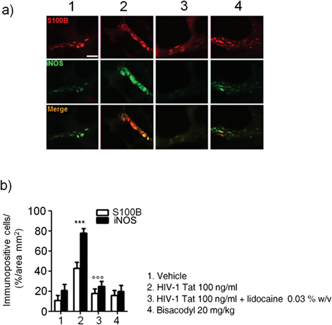
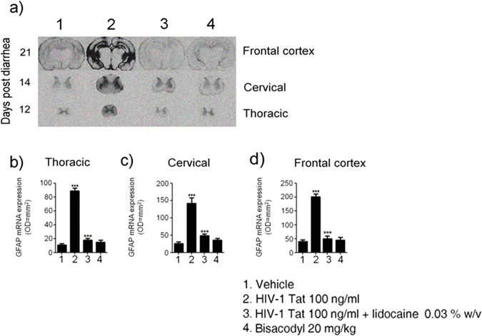
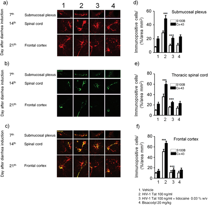
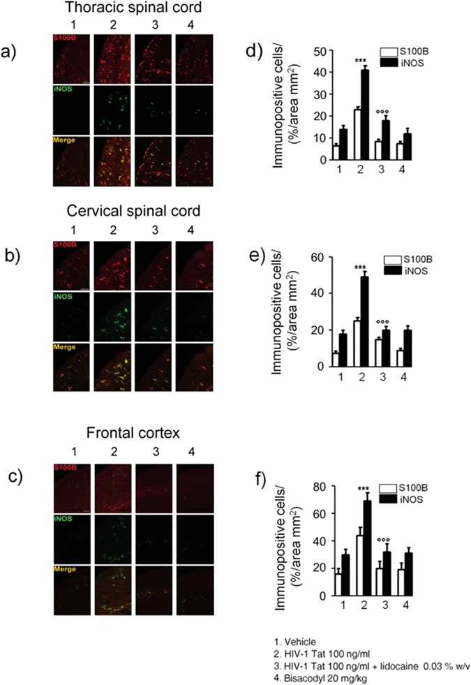
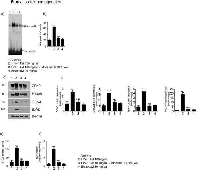
Despite the effectiveness of combined anti-retroviral therapy, human immunodeficiency virus (HIV) infected-patients frequently report diarrhea and neuropsychological deficits. It is claimed that the viral HIV-1 Trans activating factor (HIV-1 Tat) protein is responsible for both diarrhea and neurotoxic effects, but the underlying mechanisms are not known. We hypothesize that colonic application of HIV-1 Tat activates glial cells of the enteric nervous system (EGCs), leading to a neuroinflammatory response able to propagate to the central nervous system. We demonstrated that HIV-1 Tat-induced diarrhea was associated with a significant activation of glial cells within the colonic wall, the spinal cord and the frontal cortex, and caused a consistent impairment of the cognitive performances. The inhibition of glial cells activity by lidocaine, completely abolished the above-described effects. These observations point out the role of glial cells as putative effectors in HIV-1 Tat-associated gastrointestinal and neurological manifestations and key regulators of gut-brain signaling.


Conflict of interest statement

The authors declare that they have no competing interests.

Figures

Figure 1
Figure 1
Schematic representation of rats-induced diarrhea. Diagram showing the induction of diarrhea by a single intracolonic administration of HIV-1 Tat, alone or in the presence of lidocaine, or by a single dose of bisacodyl, and the time schedule for measurements on enteric, or central nervous system glia cells.
Figure 2
Figure 2
(a,b) EMSA analysis showing that intracolonic administration of HIV-1 Tat (100 ng/ml) induced a marked increase of NF-kappaB expression in EGCs nuclear extracts versus vehicle group. The administration of lidocaine significantly reduced HIV-1 Tat-induced NF-kappaB activation, whereas bisacodyl failed to induce any significant effect on NF-kappaB activation. (a) The panel shows representative NF-kappaB activation complex bands and (b) their densitometric quantification (OD = optical density in mm2). (c,d) HIV-1 Tat treatment caused a marked increase of GFAP, S100B, TLR-4 and iNOS protein expression in submucosal plexus lysates, as compared to vehicle group and this effect was significantly inhibited by lidocaine; to note that bisacodyl also failed to induce any significant effect. (c) The panel shows representative immunoreactive bands of analyzed proteins and (d) their respective levels expressed as fold change. (e,f) In the medium of submucosal plexi lysates obtained from HIV-1 Tat treated rats a significant increase of NO2 and S100B was observed in comparison with vehicle group and such effect was counteracted by lidocaine; again, bisacodyl had no effect on treated animals. Results are expressed as mean ± SEM; ***p < 0.001 vs all other groups; °°°p < 0.001 vs HIV-1 Tat group; n = 6 for each group.
Figure 3
Figure 3
Intracolonic administration of HIV-1 Tat (100 ng/ml) yields to a marked activation of submucosal plexus-EGCs, as shown by the immunofluorescence analysis showing a significant increase of S100B and iNOS protein co-expression. (a) The panel shows iNOS (green) and S100B (red) immunoreactivity with (b) the respective quantification of iNOS (filled bars) and S100B (open bars) expression in the EGCs; both lidocaine pretreatment and bisacodyl administration failed to significantly affect S100B/iNOS expression. Results are expressed as mean ± SEM; ***p < 0.001 vs all other groups; °°°p < 0.001 vs HIV-1 Tat group. Scale bar: 20 μm.
Figure 4
Figure 4
(a) In situ hybridization analysis of thoracic and cercival spinal cord, and frontal cortex showing GFAP mRNA expression at day 12, 14 and 21 after diarrhea induction, respectively. (bd) Quantitative analysis revealed that administration of HIV-1 Tat protein caused a significant increase of GFAP mRNA expression in all the analyzed areas compared, as compared to both vehicle or lidocaine group, while bisacodyl yields to no significant change. (Results are expressed as mean ± SEM; ***p < 0.001 vs all other groups; °°°p < 0.001 vs HIV-1 Tat group; OD = optical density in mm2; n = 5 for each group).
Figure 5
Figure 5
(ac) Intracolonic administration of HIV-1 Tat caused a marked increase of Cx-43 (green) protein expression in S100B-positive cells (red) in the submucosal plexus, spinal cord and frontal cortex, respectively. (df) Quantitative analysis revealed that administration of lidocaine significantly reduced Cx-43 expression on S100B-positive cells of HIV-1 Tat group, whereas bisacodyl did not produce any change in Cx-43 expression compared to vehicle group (S100B (open bars) and Cx-43 (filled bars) expression in the analyzed areas. ***p < 0.001 vs all other groups; °°°p < 0.001 vs HIV-1 Tat group. Scale bars: 20 μm; n = 6 for each group.
Figure 6
Figure 6
Intracolonic administration of HIV-1 Tat induced glial activation in the (a) thoracic and (b) cervical spinal cord and (c) frontal cortex at day 12, 14 and 21 after diarrhea induction, respectively. (ac) Immunofluorescence analysis showed that iNOS (green) and S100B (red) co-expression was increased in the spinal cord and frontal of HIV-1 Tat treated rats. (df) Quantitative analysis showed that HIV-1 Tat-induced upregulation of iNOS (filled bars) and S100B (open bars) was significantly inhibited by lidocaine treatment. Results are expressed as mean ± SEM; ***p < 0.001 vs all other groups; °°°p < 0.001 vs HIV-1 Tat group. Scale bars: 100 μm; n = 6 for each group.
Figure 7
Figure 7
Effect of HIV-1 Tat treatment on NF-kappaB activation in the nuclear extracts of frontal cortex and astrocytes activation. (a) The panel shows representative NF-kappaB activation complex bands in the different groups of rats. (b) The quantitative analysis revealed that intracolonic HIV-1 Tat administration yields to a significant increase of NF-kappaB, as compared to vehicle, lidocaine, or bisacodyl groups (OD = optical density in mm2). (c) HIV-1 Tat caused a marked increase of GFAP, S100B, TLR-4 and iNOS protein expression in the frontal cortex homogenates of treated rats. (d) Quantitative analysis reveled that HIV-1 Tat induced a significantly higher expression of GFAP, S100B, TLR-4 and iNOS, than lidocaine, or bisacodyl groups. (e,f) In the medium of frontal cortex homogenates deriving from HIV-1 Tat group a significant increase of NO2 and S100B was also observed as compared to the other groups. (Results are expressed as mean ± SEM; ***p < 0.001 vs all other groups; °°°p < 0.001 vs HIV-1 Tat group; n = 6 for each group).

References

    1. Weber, R. Enteric Infections and Diarrhea in Human Immunodeficiency Virus–Infected Persons. Arch Intern Med. 159–1473 (1999). - PubMed
    1. Goldstein G. HIV-1 Tat protein as a potential AIDS vaccine. Nat Med. 1996;1:960–964. doi: 10.1038/nm0996-960. - DOI - PubMed
    1. Simioni S, Cavassini M, Annoni J-M, et al. Cognitive dysfunction in HIV patients despite long-standing suppression of viremia. AIDS. 2010;24:1243–1250. - PubMed
    1. Bagashev A, Sawaya BE. Roles and functions of HIV-1 Tat protein in the CNS: an overview. Virol J. 2013;10:358. doi: 10.1186/1743-422X-10-358. - DOI - PMC - PubMed
    1. Canani RB, Cirillo P, Mallardo G, et al. Effects of HIV-1 Tat protein on ion secretion and on cell proliferation in human intestinal epithelial cells. Gastroenterology. 2003;124:368–76. doi: 10.1053/gast.2003.50056. - DOI - PubMed

Publication types

MeSH terms

Substances