

Acoustofluidic bacteria separation
- PMID: 28798539
- PMCID: PMC5546156
- DOI: 10.1088/1361-6439/27/1/015031
Acoustofluidic bacteria separation
Abstract
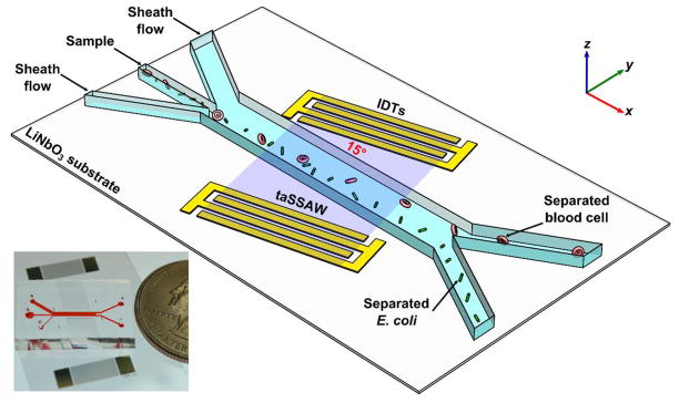
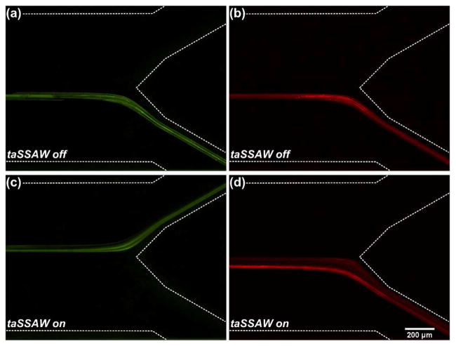
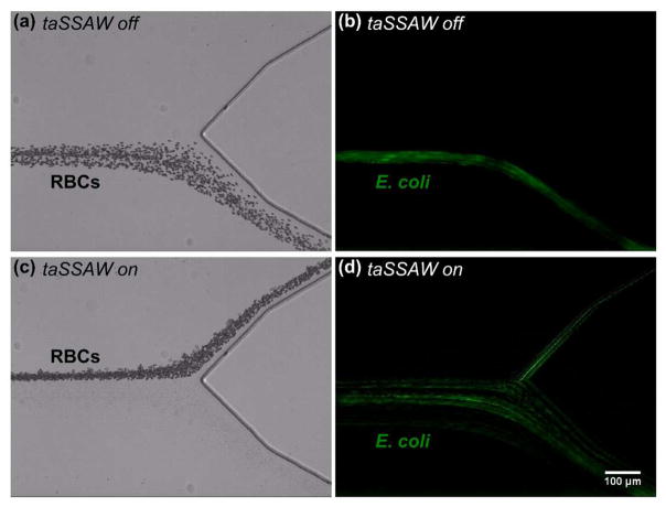
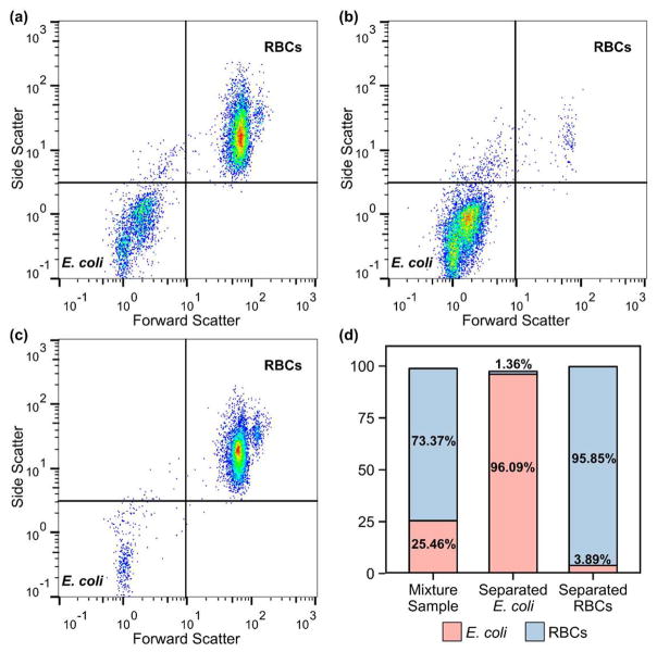
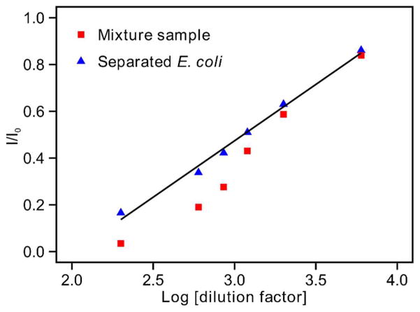
Bacterial separation from human blood samples can help with the identification of pathogenic bacteria for sepsis diagnosis. In this work, we report an acoustofluidic device for label-free bacterial separation from human blood samples. In particular, we exploit the acoustic radiation force generated from a tilted-angle standing surface acoustic wave (taSSAW) field to separate E. coli from human blood cells based on their size difference. Flow cytometry analysis of the E. coli separated from red blood cells (RBCs) shows a purity of more than 96%. Moreover, the label-free electrochemical detection of the separated E. coli displays reduced non-specific signals due to the removal of blood cells. Our acoustofluidic bacterial separation platform has advantages such as label-free separation, high biocompatibility, flexibility, low cost, miniaturization, automation, and ease of in-line integration. The platform can be incorporated with an on-chip sensor to realize a point-of-care (POC) sepsis diagnostic device.
Keywords: Acoustofluidics; bacterial separation; standing surface acoustic wave (SSAW).
Figures
References
-
- Cohen J. The immunopathogenesis of sepsis. Nature. 2002;420:885–91. - PubMed
-
- Angus DC, van der Poll T. Severe sepsis and septic shock. N Engl J Med. 2013;369:840–51. - PubMed
-
- Chaney R, Rider J, Pamphilon D. Direct detection of bacteria in cellular blood products using bacterial ribosomal RNA-directed probes coupled to electrochemiluminescence. Transfus Med. 1999;9:177–88. - PubMed
-
- Gao J, Li L, Ho P-L, Mak GC, Gu H, Xu B. Combining Fluorescent Probes and Biofunctional Magnetic Nanoparticles for Rapid Detection of Bacteria in Human Blood. Adv Mater. 2006;18:3145–8.
Grants and funding
LinkOut - more resources
Full Text Sources
Other Literature Sources