. 2017 Jul 14:11:206.
doi: 10.3389/fncel.2017.00206. eCollection 2017.

NLRP6 Inflammasome Ameliorates Brain Injury after Intracerebral Hemorrhage

Affiliations

NLRP6 Inflammasome Ameliorates Brain Injury after Intracerebral Hemorrhage

Peng-Fei Wang et al. Front Cell Neurosci. .

Abstract

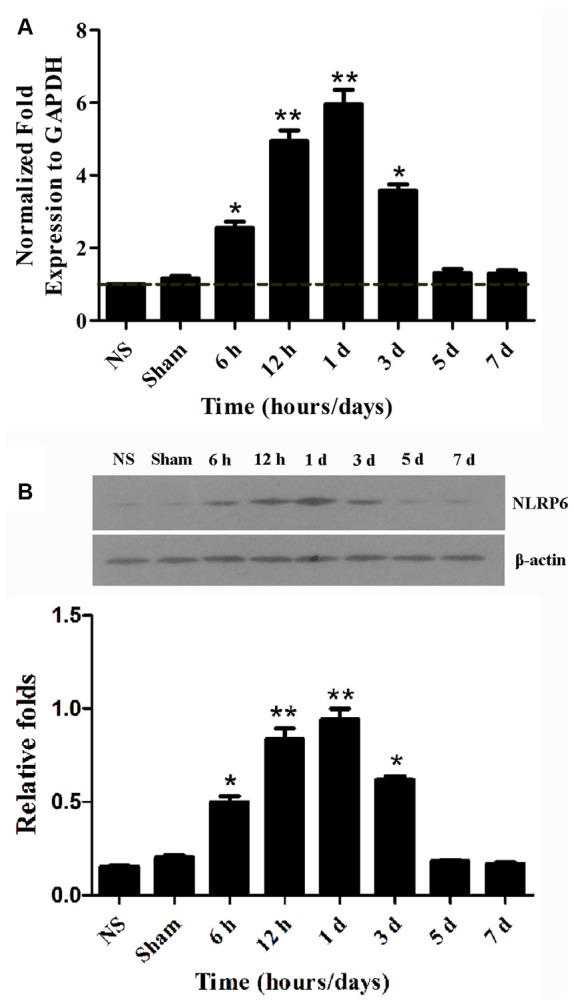
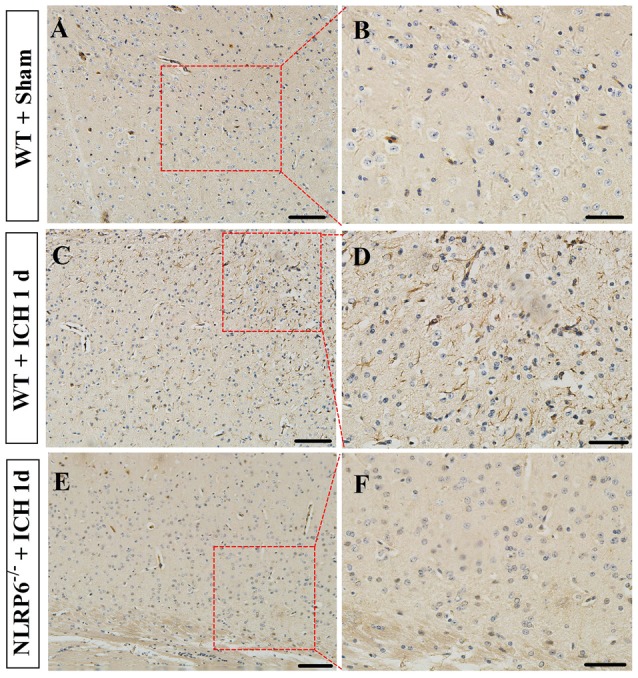
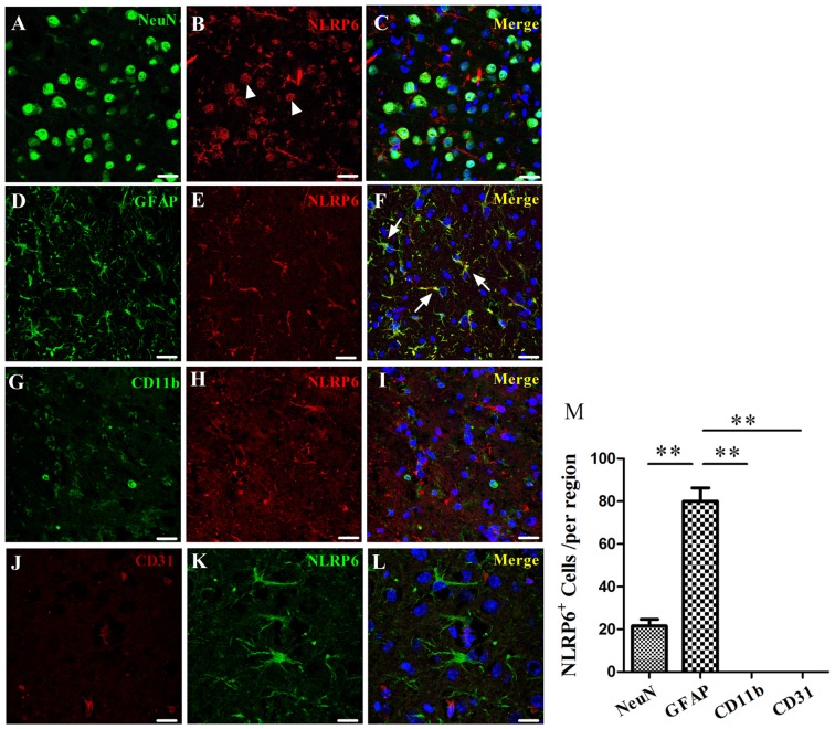
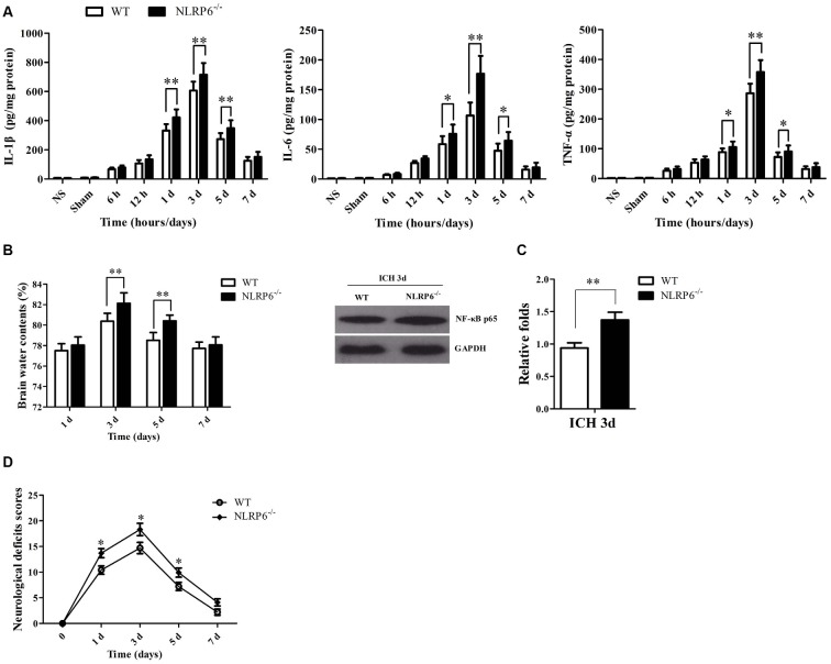
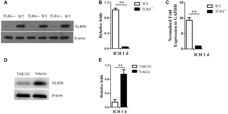
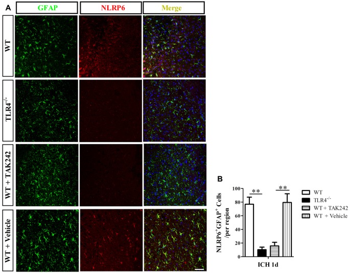
NLRP6 inflammasome, one of the important intracellular innate immune sensors, has been shown to regulate immune responses. However, its roles in the intracerebral hemorrhage (ICH) are completely not clear. In the present study, we investigated the expression profile and biological roles of NLRP6 inflammasome in perihematomal brain tissues of mice subjected to ICH. In this study, we investigated the expression profile of NLRP6 inflammasome in the perihematomal brain tissues and explored the biological role of NLRP6 inflammasome upon acute brain injury in mice subjected to ICH. Increased expression of NLRP6 inflammasome was found in perihematomal brain tissues ranging from 6 h to 3 days, with a peak level at 1 day after ICH. Immunohistochemistry staining also showed that NLRP6 inflammasome was significantly increased in the perihematomal brain tissues at 1 day after ICH. Moreover, immunofluorescence staining showed that NLRP6 inflammasome was mainly colocalized in glial fibrillary acidic protein (GFAP)-positive astrocytes, while with little colocalized expression in NeuN-positive neurons and without expression in CD11b-positive microglia and CD31-positive endothelial cell in the perihematomal brain tissue of mice after ICH. Furthermore, NLRP6-/- ICH mice exhibited significantly higher brain water contents (BMCs), proinflammatory cytokines, NF-κB activity and neurological deficit scores when compared with the wild type (WT) ICH mice. In addition, we found that Toll-like receptor 4 (TLR4)-/- mice, as well as the TAK242 treated mice, had markedly lower expression of NLRP6 inflammasome expression in the perihematomal brain tissue at 1 day after ICH. Our data suggest that the upregulated NLRP6 inflammasome in perihematomal brain tissues attenuates ICH-induced brain injury.

Keywords: NLRP6; brain injury; inflammasome; intracerebral hemorrhage; neuroinflammation.


Figures

Figure 1
Figure 1
NLRP6 expression in the perihematomal brain tissues after intracerebral hemorrhage (ICH). (A) Real-time PCR results showing NLRP mRNA expression in the perihematomal brain tissues of mice at 6 h to 7 days post-ICH. (B) Representative bands and densitometric quantification of NLRP6 expression in the perihematomal brain tissues of mice at 6 h to 7 days post-ICH. n = 6 per group, **P < 0.01 vs. the normal brain (NS) and Sham groups; *P < 0.05 vs. the NS and Sham groups.
Figure 2
Figure 2
Immunohistochemical staining for NLRP6. (A,B) Representative images of NLRP6 immunostaining in the brain tissues in the Sham groups. (C,D) Representative images of NLRP6 immunostaining in the perihematomal brain tissues at 1 day after ICH. (E,F) Representative images of NLRP6 immunostaining in the brain tissues in the NLRP6−/− mice at 1 day after ICH. n = 4 per group, scale bars = 100 μm (A,C,E), scale bars = 50 μm (B,D,F).
Figure 3
Figure 3
Immunofluorescence staining for NLRP6 after ICH. (A–C) Immunofluorescence staining for NeuN (A) and NLRP6 (B); the merged image shows that little NLRP6 was expressed in NeuN-positive neurons (C; arrow head). (D–F) Immunofluorescence staining for glial fibrillary acidic protein (GFAP) (D) and NLRP6 (E); the merged image shows that NLRP6 staining was also observed in the cytoplasm and end processes of astrocytes (F; arrow). (G–I) Immunofluorescence staining for CD11b (G) and NLRP6 (H); the merged image shows that NLRP6 was not expressed in CD11b-positive microglia (I). (J–L) Immunofluorescence staining for CD31 (J) and NLRP6 (K); the merged image shows that NLRP6 was not expressed in CD31-positive endothelial cell (L). All images were captured in the perihematomal brain tissues. (M) Densitometric quantification of NLRP6 expression in the perihematomal brain tissues at 1 day after ICH (n = 4 per group, **P < 0.01). Scale bars = 25 μm (A–I); = 15 μm (J–L).
Figure 4
Figure 4
The absence of NLRP6 inflammasome significantly deteriorate the brain injury caused ICH. (A) Levels of inflammatory factors in perihematomal brain tissues of mice in the normal brain, sham, 6 h, 12 h, 1 day, 3 days, 5 days and 7 days after ICH (n = 6 per group, **P < 0.01; *P < 0.05). (B) Brain water content (BMC) in the wild type (WT) and NLRP6−/− mice at 1 day, 3 days, 5 days and 7 days after ICH (n = 6 per group, **P < 0.01). (C) Representative bands and densitometric quantification of NF-κB p65 expression in the perihematomal brain tissues at 1 day post-ICH between WT and NLRP6−/− mice (n = 4 per group, **P < 0.01). (D) Neurological Deficient Score (NDS) in the WT and NLRP6−/− mice at 1 day, 3 days, 5 days and 7 days after ICH (n = 9 per group, *P < 0.05 vs. the WT group).
Figure 5
Figure 5
Increased NLRP6 inflammasome expression may be involved in Toll-like receptor 4 (TLR4) signaling after ICH. (A) Representative western blot results showing NLRP6 expression between WT and TLR4−/− mice at 1 day after ICH. (B) Densitometric quantification of NLRP6 expression in the perihematomal brain tissues of WT and TLR4−/− mice at 1 day after ICH (n = 6, **P < 0.01). (C) Real-time PCR results showing NLRP6 mRNA expression (n = 5, **P < 0.01). (D) Representative western blot results showing NLRP6 expression between TAK242 and Vehicle treated mice at 1 day after ICH. (E) Densitometric quantification of NLRP6 expression in the perihematomal brain tissues of TAK242 and Vehicle treated mice at 1 day after ICH (n = 4, **P < 0.01).
Figure 6
Figure 6
Immunofluorescence staining for NLRP6 inflammasome in TAK242 treated ICH mice. Representative (A) and statistical graph (B) of immunofluorescent staining of cells expressing NLRP6 and GFAP by astrocytes 1 day after ICH among WT, TLR4−/−, WT + TAK242 and WT + Vehicle mice (n = 4, **P < 0.01). Scale bars = 50 μm.

References

    1. Abulafia D. P., de Rivero Vaccari J. P., Lozano J. D., Lotocki G., Keane R. W., Dietrich W. D. (2009). Inhibition of the inflammasome complex reduces the inflammatory response after thromboembolic stroke in mice. J. Cereb. Blood Flow Metab. 29, 534–544. 10.1038/jcbfm.2008.143 - DOI - PubMed
    1. Akira S., Takeda K. (2004). Toll-like receptor signalling. Nat. Rev. Immunol. 4, 499–511. 10.1038/nri1391 - DOI - PubMed
    1. Anand P. K., Malireddi R. K., Lukens J. R., Vogel P., Bertin J., Lamkanfi M., et al. . (2012). NLRP6 negatively regulates innate immunity and host defence against bacterial pathogens. Nature 488, 389–393. 10.1038/nature11250 - DOI - PMC - PubMed
    1. Chen G. Y., Liu M., Wang F., Bertin J., Núñez G. (2011). A functional role for Nlrp6 in intestinal inflammation and tumorigenesis. J. Immunol. 186, 7187–7194. 10.4049/jimmunol.1100412 - DOI - PMC - PubMed
    1. Elinav E., Strowig T., Kau A. L., Henao-Mejia J., Thaiss C. A., Booth C. J., et al. . (2011). NLRP6 inflammasome regulates colonic microbial ecology and risk for colitis. Cell 145, 745–757. 10.1016/j.cell.2011.04.022 - DOI - PMC - PubMed

LinkOut - more resources