. 2017 Jul 17;11(4):044104.
doi: 10.1063/1.4991738. eCollection 2017 Jul.

The role of shear stress and altered tissue properties on endothelial to mesenchymal transformation and tumor-endothelial cell interaction

Affiliations

The role of shear stress and altered tissue properties on endothelial to mesenchymal transformation and tumor-endothelial cell interaction

Sara G Mina et al. Biomicrofluidics. .

Abstract

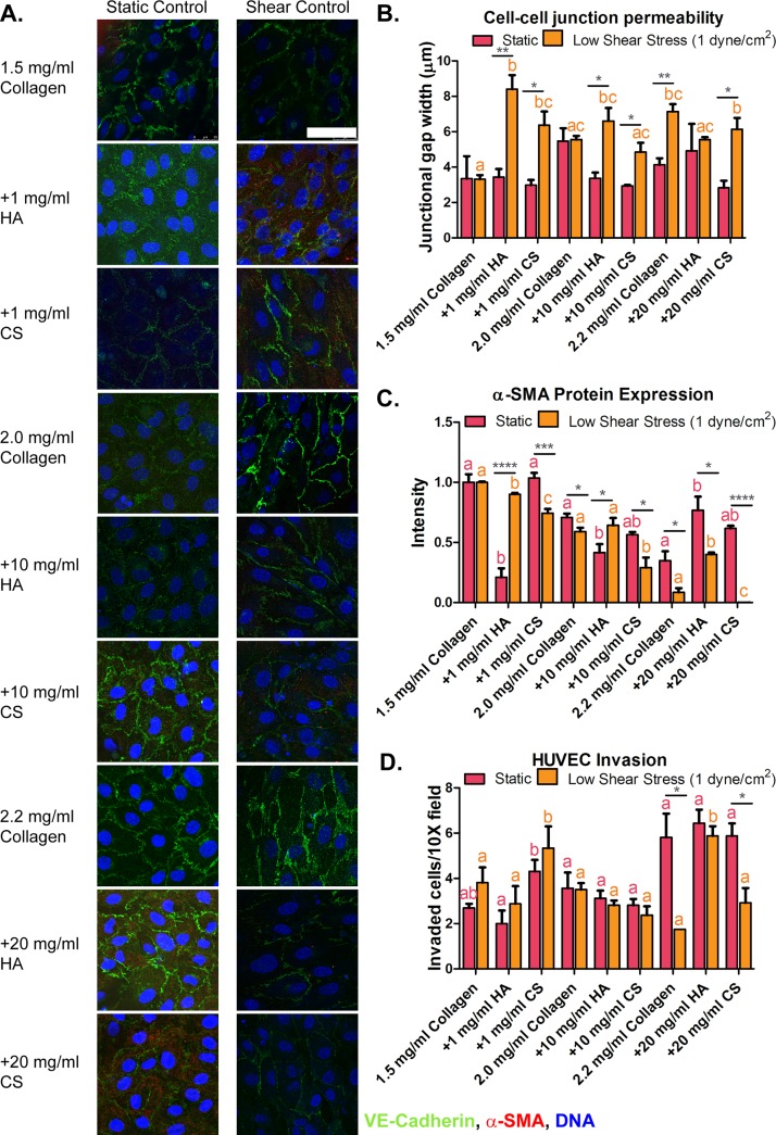
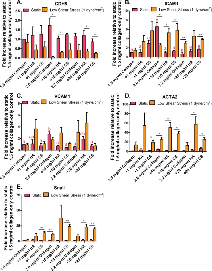
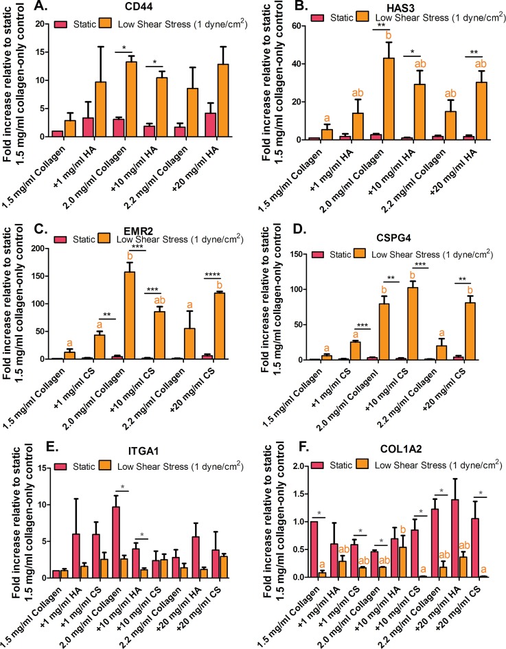
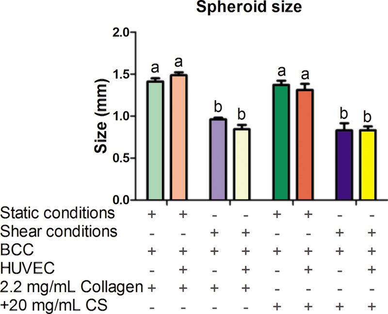
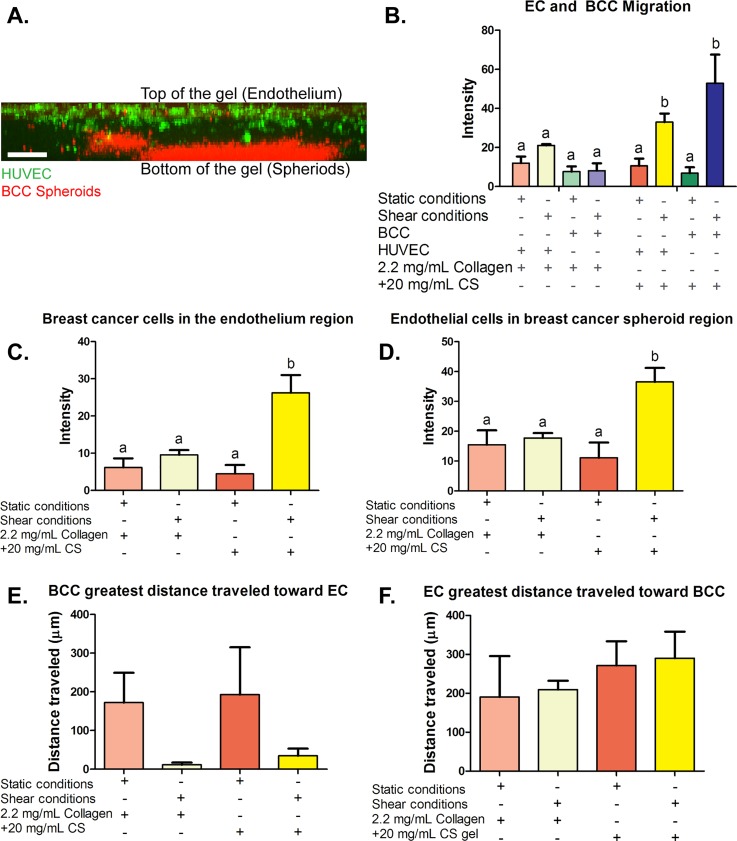
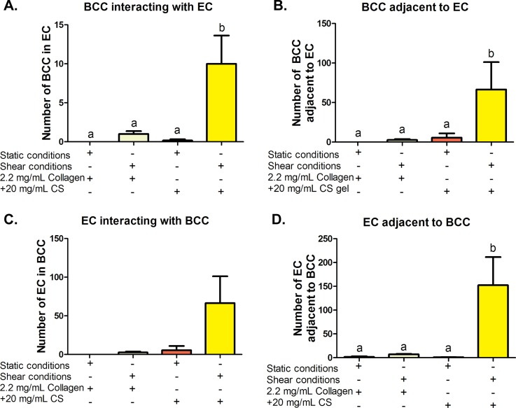
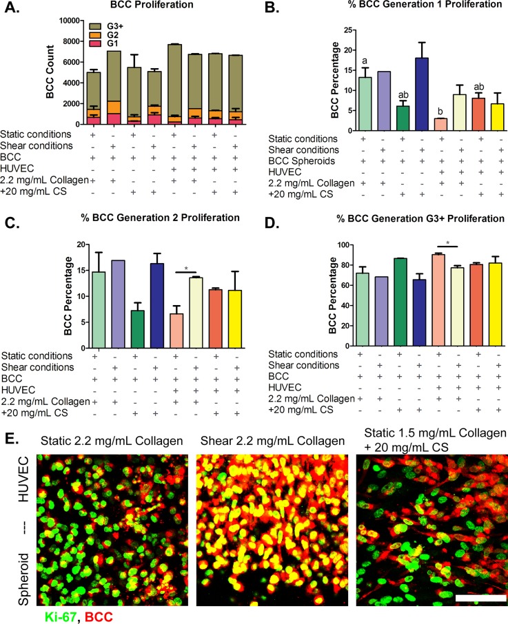
Tumor development is influenced by stromal cells in aspects including invasion, growth, angiogenesis, and metastasis. Activated fibroblasts are one group of stromal cells involved in cancer metastasis, and one source of activated fibroblasts is endothelial to mesenchymal transformation (EndMT). EndMT begins when the endothelial cells delaminate from the cell monolayer, lose cell-cell contacts, lose endothelial markers such as vascular endothelial-cadherin (VE-cadherin), gain mesenchymal markers like alpha-smooth muscle actin (α-SMA), and acquire mesenchymal cell-like properties. A three-dimensional (3D) culture microfluidic device was developed for investigating the role of steady low shear stress (1 dyne/cm2) and altered extracellular matrix (ECM) composition and stiffness on EndMT. Shear stresses resulting from fluid flow within tumor tissue are relevant to both cancer metastasis and treatment effectiveness. Low and oscillatory shear stress rates have been shown to enhance the invasion of metastatic cancer cells through specific changes in actin and tubulin remodeling. The 3D ECM within the device was composed of type I collagen and glycosaminoglycans (GAGs), hyaluronic acid and chondroitin sulfate. An increase in collagen and GAGs has been observed in the solid tumor microenvironment and has been correlated with poor prognosis in many different cancer types. In this study, it was found that ECM composition and low shear stress upregulated EndMT, including upregulation of mesenchymal-like markers (α-SMA and Snail) and downregulated endothelial marker protein and gene expression (VE-cadherin). Furthermore, this novel model was utilized to investigate the role of EndMT in breast cancer cell proliferation and migration. Cancer cell spheroids were embedded within the 3D ECM of the microfluidic device. The results using this device show for the first time that the breast cancer spheroid size is dependent on shear stress and that the cancer cell migration rate, distance, and proliferation are induced by EndMT-derived activated fibroblasts. This model can be used to explore new therapeutics in a tumor microenvironment.


Figures

FIG. 1.
FIG. 1.
Protein expression and invasion of human umbilical vein endothelial cells (HUVEC) seeded onto 3D hydrogels. (a) HUVEC immunocytochemistry images for alpha-smooth muscle actin (α-SMA; red), vascular endothelial-cadherin (VE-cadherin; green), and cell DNA (blue) on three-dimensional (3D) collagen-only or collagen + glycosaminoglycans [GAGs, chondroitin sulfate (CS) or hyaluronic acid (HA)] gels after 48 h of exposure to static and flow-induced 1 dyne/cm2 shear stress. (b) Gap junction width measurements of HUVEC seeded onto 3D collagen-only or collagen + GAGs hydrogels exposed to static and 1 dyne/cm2 shear stress conditions. (c) Fluorescence intensity analysis of mesenchymal marker (α-SMA) protein expression in HUVEC. (d) HUVEC invasion into the extracellular matrix of cells cultured on collagen-only controls and collagen-GAG exposed to static conditions and 1 dyne/cm2 shear stress. Fluorescence intensity analysis of mesenchymal marker α-SMA protein expression in HUVEC. Data shown are mean ± SEM, n ≥ 3 culture wells. All static or all fluidic conditions were analyzed with a one-way ANOVA with Tukey's post-test. Tukey groups are shown for statistically significant results (p < 0.05). Identical extracellular matrix conditions exposed to static or shear conditions were analyzed with an unpaired Student's t-test. Bars connected with * represent statistical significance with an unpaired Student's t-test (p < 0.05). Scale bar = 50 μm.
FIG. 2.
FIG. 2.
Human umbilical vein endothelial cells (HUVEC) inflammatory and pro-EndMT gene expression. (a) Vascular endothelial-cadherin (VE-cadherin; CDH5), (b) pro-inflammatory cell receptor intercellular adhesion molecule-1 (ICAM-1), and (c) vascular cell adhesion molecule-1 (VCAM-1). (d) Endothelial to mesenchymal transformation (EndMT) markers, actin, alpha 2 (ACTA2) and (e) Snail. Error bars show mean ± SEM, n = 3 culture wells. All static or all fluidic conditions were analyzed with a one-way ANOVA with Tukey's post-test. Tukey groups are shown for statistically significant results (p < 0.05). Identical extracellular matrix conditions exposed to static or shear conditions were analyzed with an unpaired Student's t-test. Bars connected with * represent statistical significance with an unpaired Student's t-test (p < 0.05).
FIG. 3.
FIG. 3.
Human umbilical vein endothelial cells (HUVEC) glycosaminoglycan (GAG) and collagen cell-surface receptors and extracellular matrix (ECM) remodeling gene expression. (a) Cell-surface receptor for hyaluronic acid (HA; CD44) and (b) production of HA (HAS3). (c) Cell-surface receptor for chondritic sulfate (CS; EMR2) and (d) production of CS (CSPG4). (e) Collagen 1 subunit integrin receptor (ITGA1) and (f) fibrosis marker (COL1A2). Error bars show mean ± SEM, n = 3 culture wells. All static or all fluidic conditions were analyzed with a one-way ANOVA with Tukey's post-test. Tukey groups are shown for statistically significant results (p < 0.05). Identical extracellular matrix conditions exposed to static or shear conditions were analyzed with an unpaired Student's t-test. Bars connected with * represent statistical significance with an unpaired Student's t-test (p < 0.05).
FIG. 4.
FIG. 4.
Breast cancer spheroid size. Spheroid size was measured following embedding in three-dimensional (3D) collagen-only or collagen + chondroitin sulfate (CS) gels and after 48 h of exposure to static or shear stress conditions. Error bars show mean ± SEM, n ≥ 8 spheroids and n = 2 biological samples. Bars that do not share any letters are significantly different according to a two-way ANOVA with Tukey's post-hoc test (p < 0.05).
FIG. 5.
FIG. 5.
Migration and migration distance quantification of breast cancer cells (BCC) and human umbilical vein endothelial cells (HUVEC). (a) Side view confocal image of the three-dimensional (3D) hydrogels with live cell staining. Live cell stain of human umbilical vein endothelial cells (HUVEC) on top of the gels (green) and live cell staining of spheroids (red) embedded in a collagen hydrogel. (b) BCC embedded in three-dimensional (3D) collagen-only gels and collagen +20 mg/ml chondroitin sulfate (CS) gels after 48 h of exposure to static or shear stress conditions migration toward the endothelium. (c) Fluorescent intensity quantification of BCC stained with CellTrace Far Red in the endothelium region and (d) fluorescent intensity quantification of EC stained with CellTrace Green in the spheroid region. (e) and (f) Greatest distance breast cancer and endothelial cells traveled in 2.2. mg/ml collagen-only or collagen + 20 mg/ml chondroitin sulfate (CS) gels after 48 h of exposure to static or shear stress conditions. (e) Quantification of the greatest distance BCC traveled. (f) Quantification of the greatest distance HUVEC traveled. Error bars show mean ± SEM, n = 6 confocal images from 2 biological samples. All conditions were analyzed with a one-way ANOVA with Tukey's post-test. Tukey groups are shown for statistically significant results (p < 0.05). Scale bar = 250 μm.
FIG. 6.
FIG. 6.
Quantification of breast cancer cell (BCC)-human umbilical vein endothelial cell (HUVEC) interactions. (a) The number of BCC inside a group of HUVEC and (b) physically touching HUVEC and likewise (c) HUVEC count inside a group of BCC and (d) number of HUVEC touching BCC. Error bars show SEM, n = 6 confocal images from 2 biological samples. Bars that do not share any letters are significantly different according to a two-way ANOVA with Tukey's post-hoc test (p < 0.05).
FIG. 7.
FIG. 7.
Breast cancer cells (BCC) proliferation. BCC were cultured for 48 h with human umbilical vein endothelial cells in three-dimensional collagen or collagen + chondroitin sulfate (CS) gels for 48 h under static or shear conditions. (a) Total cell count for all generations. Cell generations are noted as (b) G1, (c) G2, and (d) G3 and higher. (e) Ki-67 proliferation marker immunocytochemistry confocal microscopy images of BCC. BCC spheroids embedded in 2.2 mg/ml collagen-only stiffness control cultured with human umbilical vein endothelial cells (HUVEC) on top of the gels and exposed to (Left) static conditions and (Middle) shear stress conditions. (Right) BCC spheroids embedded in 1.5 mg/ml collagen + 20 mg/ml CS cultured with HUVEC on top of the gels and exposed to static conditions. BCC were stained with CellTrace Far Red and Ki-67 proliferation marker (green). Bar Scale = 50 μm. Error bars show mean ± SEM, n = 2 biological samples. All static or all fluidic conditions were analyzed with a one-way ANOVA with Tukey's post-test. Tukey groups are shown for statistically significant results (p < 0.05). Identical extracellular matrix conditions exposed to static or shear conditions were analyzed with an unpaired Student's t-test. Bars connected with * represent statistical significance with an unpaired Student's t-test (p < 0.05). Scale bar = 50 μm.

Similar articles

Cited by

References

    1. Weir H. K., Thompson T. D., Soman A., Moller B., and Leadbetter S., Cancer 121, 1827 (2015).10.1002/cncr.29258 - DOI - PMC - PubMed
    1. Bray F. and Møller B., Nat. Rev. Cancer 6, 63 (2006).10.1038/nrc1781 - DOI - PubMed
    1. Mariotto A. B., Yabroff K. R., Shao Y., Feuer E. J., and Brown M. L., J. Natl. Cancer Inst. 103, 117 (2011).10.1093/jnci/djq495 - DOI - PMC - PubMed
    1. Schroer A. K. and Merryman W. D., J. Cell Sci. 128, 1865 (2015).10.1242/jcs.162891 - DOI - PMC - PubMed
    1. Frangogiannis N. G., Nat. Rev. Cardiol. 11, 255 (2014).10.1038/nrcardio.2014.28 - DOI - PMC - PubMed

LinkOut - more resources