Review
. 2017 Jul 26:5:40.
doi: 10.3389/fbioe.2017.00040. eCollection 2017.

Tissue Engineering Approaches in the Design of Healthy and Pathological In Vitro Tissue Models

Affiliations
Review

Tissue Engineering Approaches in the Design of Healthy and Pathological In Vitro Tissue Models

Silvia Caddeo et al. Front Bioeng Biotechnol. .

Abstract

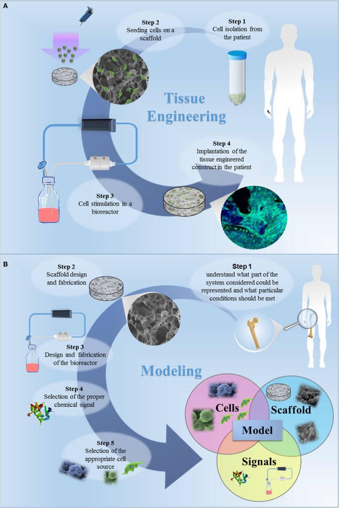
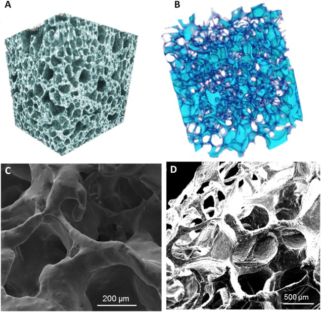
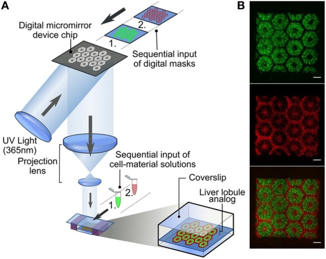
In the tissue engineering (TE) paradigm, engineering and life sciences tools are combined to develop bioartificial substitutes for organs and tissues, which can in turn be applied in regenerative medicine, pharmaceutical, diagnostic, and basic research to elucidate fundamental aspects of cell functions in vivo or to identify mechanisms involved in aging processes and disease onset and progression. The complex three-dimensional (3D) microenvironment in which cells are organized in vivo allows the interaction between different cell types and between cells and the extracellular matrix, the composition of which varies as a function of the tissue, the degree of maturation, and health conditions. In this context, 3D in vitro models can more realistically reproduce a tissue or organ than two-dimensional (2D) models. Moreover, they can overcome the limitations of animal models and reduce the need for in vivo tests, according to the "3Rs" guiding principles for a more ethical research. The design of 3D engineered tissue models is currently in its development stage, showing high potential in overcoming the limitations of already available models. However, many issues are still opened, concerning the identification of the optimal scaffold-forming materials, cell source and biofabrication technology, and the best cell culture conditions (biochemical and physical cues) to finely replicate the native tissue and the surrounding environment. In the near future, 3D tissue-engineered models are expected to become useful tools in the preliminary testing and screening of drugs and therapies and in the investigation of the molecular mechanisms underpinning disease onset and progression. In this review, the application of TE principles to the design of in vitro 3D models will be surveyed, with a focus on the strengths and weaknesses of this emerging approach. In addition, a brief overview on the development of in vitro models of healthy and pathological bone, heart, pancreas, and liver will be presented.

Keywords: bone; heart; liver; models; pancreas; three-dimensional; tissue engineering.


Figures

Figure 1
Figure 1
(A) The classic tissue engineering (TE) paradigm consists in the isolation of primary cell from the patient and the further seeding on a three-dimensional (3D) porous scaffold, specifically designed to induce cell proliferation and differentiation. The bioartificial system is developed in a specific environment, where the metabolic, mechanical and electrical stimuli are provided by a bioreactor. In this tissue-specific designed system, cells start to produce extracellular matrix, leading to in vitro tissue maturation. The tissue-engineered construct is then implanted in the patient, undergoing to a remodeling process in vivo, which allows the tissue restoration. (B) The TE approach for the development of in vitro models. The first step is to understand what part of the system considered should be represented and which specific conditions should be met. The second step is the design of the appropriate scaffold, i.e., the design of a 3D porous construct recapitulating the architecture and the mechanical and surface properties of the tissue/organ to be modeled. The third step is the selection of the cells to be seeded in the designed scaffold. The subsequent steps should be the design and fabrication of a bioreactor and the selection of the appropriate chemical signals (e.g., cocktail of growth factors) to be included in the model. Once a model is successfully developed, it can be improved by increasing its complexity, by introducing additional cell type, chemical factor or coupling different mechanical stimuli. The final step is the validation, aiming to demonstrate the relevance of the model for the intended purpose. The validation procedure is fundamental and this step should be carefully designed.
Figure 2
Figure 2
Micro-CT reconstruction of an example of glass–ceramic scaffolds mimicking (A) healthy bone and (B) osteoporotic bone architectures. SEM micrographs of (C) a “healthy” scaffold and (D) an “osteoporotic” scaffold. Magnifications: (C) 300×, (D) 100×. Reprinted with permission from Baino et al. (2016).
Figure 3
Figure 3
Human iPSCs (hiPSCs) were obtained by reprogramming dermal fibroblasts. Upon differentiation toward the cardiac phenotype (hiPSC-CMs), the two-dimensional cardiac tissue model was designed by plating the cells onto fibronectin-coated glass coverslips. (A) hiPSC-CMs suffering long QT syndrome (LQTS) stained for α-actinin, troponin I (TnI) and connexion 43 (Cx43). White arrows identify gap junctions Scale bars: 30 µm. (B) Polymerase chain reaction results showing that both LQTS and control hiPSC-CMs express cardiac-specific transcription factors (NKX2-5), sarcomeric proteins (MYH6, MYH7, MLC2V), and ion channels (KCNH2), while undifferentiated hiPSCs express the pluripotency markers OCT4 and NANOG. (C) Extracellular recordings and field-potential duration (FPD) measurements from healthy hiPSC-derived cardiac tissue (control) (top) and hiPSC-derived cardiac tissue suffering LQTS. FPD was significantly longer in cardiac tissues derived from LQTS hiPSC-CMs in comparison to the control. (D) LQTS cardiac tissue from hiPSC-CMs showed marked arrhythmogenicity. Arrows identify single and multiple premature beats (Itzhaki et al., 2011).
Figure 4
Figure 4
Schematic reproduction of the microbeads’ microfluidic synthesis, and three-dimensional (3D) cell culture. The microbeads were made from cross-linked poly(ethylene glycol)-co-poly-l-lysine (PEG-co-PLL) hydrogels modified with the cell surface receptor and its membrane-bound ligand pair, EphA/EphrinA, and coated with pancreatic tissue-specific extracellular matrix components derived from rat pancreatic decellularized matrix. The microbeads (in red) were then cultured in direct contact with β cells (in green) mimicking the cell–cell interaction that cell experiences in vivo. Reprinted with permission from Li et al. (2013a).
Figure 5
Figure 5
Three-dimensional (3D) printed hepatic lobule models. (A) 3D bioprinting of human iPSCs (hiPSC)-derived hepatocytes and supporting cells encapsulated in photocurable hydrogels [5%w/v gelatin methacrylate (GelMa) for hiPSC-derived hepatocytes to obtain, after photopolymerization a matrix with stiffness similar to the healthy liver tissue, a blend of glycidal methacrylate-hyaluronic acid and GelMa for supporting endothelial and mesenchymal cells to favor endothelial cell proliferation and support vascularization processes] was carried out in two steps according to well-defined patterns (B) to finely reproduce their localization in native lobules. In detail, hiPSC-derived hepatocytes were first patterned according to a digital mask with proper geometry, followed by the patterning of the supporting cells using a second digital mask (scale bars 500 µm, supporting cells and hiPSC-hepatocytes were fluorescently marked in red and green, respectively). Reprinted with permission from Ma et al. (2016).

Similar articles

Cited by

References

    1. Adams C. P., Brantner V. V. (2006). Estimating the cost of new drug development: is it really $802 million? Health Aff. 25, 420–428.10.1377/hlthaff.25.2.420 - DOI - PubMed
    1. Aerssens J., Boonen S., Lowet G., Dequeker J. (1998). Interspecies differences in bone composition, density, and quality: potential implications for in vivo bone research 1. Endocrinology 139, 663–670.10.1210/endo.139.2.5751 - DOI - PubMed
    1. Alvarez K., Nakajima H. (2009). Metallic scaffolds for bone regeneration. Materials 2, 790–832.10.3390/ma2030790 - DOI
    1. American Diabetes Association. (2010). Diagnosis and classification of diabetes mellitus. Diabetes Care 33(Suppl. 1), S62–S69.10.2337/dc10-S062 - DOI - PMC - PubMed
    1. Andersson A. (1978). Isolated mouse pancreatic islets in culture: effects of serum and different culture media on the insulin production of the islets. Diabetologia 14, 397–404.10.1007/BF01228134 - DOI - PubMed

LinkOut - more resources