. 2017:2017:5806832.
doi: 10.1155/2017/5806832. Epub 2017 Jul 17.

Lycium barbarum Polysaccharides Protect Rat Corneal Epithelial Cells against Ultraviolet B-Induced Apoptosis by Attenuating the Mitochondrial Pathway and Inhibiting JNK Phosphorylation

Affiliations

Lycium barbarum Polysaccharides Protect Rat Corneal Epithelial Cells against Ultraviolet B-Induced Apoptosis by Attenuating the Mitochondrial Pathway and Inhibiting JNK Phosphorylation

Shaobo Du et al. Biomed Res Int. 2017.

Abstract

Lycium barbarum polysaccharides (LBPs) have been shown to play a key role in protecting the eyes by reducing the apoptosis induced by certain types of damage. However, it is not known whether LBPs can protect damaged corneal cells from apoptosis. Moreover, no reports have focused on the role of LBPs in guarding against ultraviolet B- (UVB-) induced apoptosis. The present study aimed to investigate the protective effect and underlying mechanism of LBPs against UVB-induced apoptosis in rat corneal epithelial (RCE) cells. The results showed that LBPs significantly prevented the loss of cell viability and inhibited cell apoptosis induced by UVB in RCE cells. LBPs also inhibited UVB-induced loss of mitochondrial membrane potential, downregulation of Bcl-2, and upregulation of Bax and caspase-3. Finally, LBPs attenuated the phosphorylation of c-Jun NH2-terminal kinase (JNK) triggered by UVB. In summary, LBPs protect RCE cells against UVB-induced damage and apoptosis, and the underlying mechanism involves the attenuation of the mitochondrial apoptosis pathway and the inhibition of JNK phosphorylation.


Figures

Figure 1
Figure 1
Schematic diagram showing the procedure for the experimental model corresponding to the LBP treatment group (UVB+/LBPs+).
Figure 2
Figure 2
Effects of LBPs on the cell viability of RCE cells. Cells were incubated for 24 h in different concentrations of LBPs. Data are presented as mean ± standard deviation, n = 3. ∗∗P < 0.01 compared with 0 mg/mL LBPs.
Figure 3
Figure 3
LBPs prevented the UVB-induced decline in cell viability in RCE cells. Data are presented as mean ± standard deviation, n = 3. aP < 0.01 compared with the UVB−/LBPs− group; bP < 0.01 compared with the UVB+/LBPs− group; cP < 0.05 compared with the UVB+/LBPs− group. ns, no significant difference.
Figure 4
Figure 4
Inhibitory effect of 1 mg/mL LBPs on UVB-induced apoptosis in RCE cells. (a) Flow cytometry scatter plot, including UVB−/LBPs−, UVB−/LBPs+, UVB+/LBPs−, and UVB+/LBPs+ groups. Q1: dead cells; Q2: late apoptosis cells; Q3: early apoptosis cells; Q4: normal cells. (b) Histogram of the quantitative change in apoptotic rates. Apoptotic rate = early cellular apoptotic rate + late cellular apoptotic rate. Data are presented as mean ± standard deviation, n = 3. P < 0.05 and ∗∗P < 0.01. (c) Changes in cellular morphology of RCE cells in each group.
Figure 5
Figure 5
Relative fluorescence intensities of rhodamine 123, showing that LBPs prevent UVB-induced MMP loss. Data are presented as mean ± standard deviation, n = 3. P < 0.05 compared with the UVB−/LBPs− group; #P < 0.05 compared with the UVB+/LBPs− group.
Figure 6
Figure 6
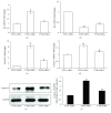
LBPs inhibit UVB-induced upregulation of Bax and caspase-3 and downregulation of Bcl-2 in RCE cells. (a, b) Relative mRNA expression changes of Bax and Bcl-2 in UVB−/LBPs−, UVB+/LBPs−, and UVB+/LBPs+ groups. (c) Fold changes in the Bax/Bcl-2 ratio in each group. (d) Relative mRNA expression changes of caspase-3 in each group. (e) Changes in the protein levels of caspase-3 were confirmed by Western blotting. GAPDH was used as an endogenous control protein. (f) Fold changes in the relative optical intensities of caspase-3/GAPDH in each group. P < 0.05 and ∗∗P < 0.01 compared with the UVB−/LBPs− group; #P < 0.05 and ##P < 0.01 compared with the UVB+/LBPs− group; n = 3 in each group.
Figure 7
Figure 7
LBPs attenuate the phosphorylation of JNK triggered by UVB in RCE cells. (a) Both p-JNK and nonphosphorylated JNK were detected by Western blotting. GAPDH was used as an endogenous control protein. (b) Fold changes of the relative optical intensities of p-JNK/JNK. P < 0.05 compared with the UVB−/LBPs− group; #P < 0.05 compared with the UVB+/LBPs− group; n = 3 in each group.

Similar articles

Cited by

References

    1. Kolozsvari L., Nogradi A., Hopp B., Bor Z. UV absorbance of the human cornea in the 240- to 400-nm range. Investigative Ophthalmology & Visual Science. 2002;43(7):2165–2168. - PubMed
    1. Downes J. E., Swann P. G., Holmes R. S. Differential corneal sensitivity to ultraviolet light among inbred strains of mice: Correlation of ultraviolet B sensitivity with aldehyde dehydrogenase deficiency. Cornea. 1994;13(1):67–72. doi: 10.1097/00003226-199401000-00011. - DOI - PubMed
    1. Ardan T., Němcová L., Bohuslavová B., et al. Reduced Levels of Tissue Inhibitors of Metalloproteinases in UVB-Irradiated Corneal Epithelium. Photochemistry and Photobiology. 2016;92(5):720–727. doi: 10.1111/php.12612. - DOI - PubMed
    1. Youn H.-Y., McCanna D. J., Sivak J. G., Jones L. W. In vitro ultraviolet-induced damage in human corneal, lens, and retinal pigment epithelial cells. Molecular Vision. 2011;17:237–246. - PMC - PubMed
    1. Balaiya S., Murthy R. K., Brar V. S., Chalam K. V. Evaluation of ultraviolet light toxicity on cultured retinal pigment epithelial and retinal ganglion cells. Clinical Ophthalmology. 2010;4(1):33–39. - PMC - PubMed

MeSH terms

Substances

LinkOut - more resources