

A Fatal Case of Erdheim-Chester Disease with Hepatic Involvement
- PMID: 28798943
- PMCID: PMC5541758
- DOI: 10.14309/crj.2017.95
A Fatal Case of Erdheim-Chester Disease with Hepatic Involvement
Abstract
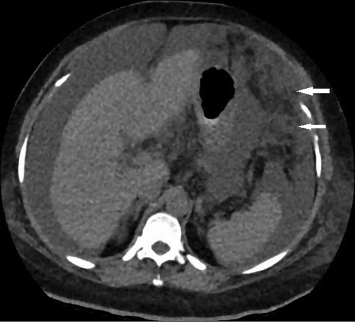
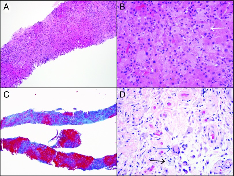
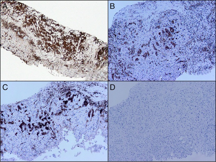
Erdheim-Chester disease (ECD) is a rare form of systemic histiocytosis, typically presenting with striking osseous involvement characterized by bilateral osteosclerosis and involvement of organs such as the lung, pituitary gland, heart, and brain. Liver involvement with ECD is extremely uncommon. We report a 56-year-old woman presenting with newly diagnosed cirrhosis and signs concerning for intra-abdominal malignancy, including omental caking and peritoneal thickening. Liver biopsy demonstrated xanthogranulomatous infiltration from ECD. The patient showed initial improvement with interferon therapy, but she developed severe depression, which led to the discontinuation of the treatment. Shortly afterward, she died from progressive liver dysfunction resulting in hepatorenal syndrome.
Figures
References
-
- Chester W. Uber lipoidgranulomatose. Virchows Arch Pathol Anat. 1930;279:561–602.
-
- Cavalli G, Guglielmi B, Berti A, et al. The multifaceted clinical presentations and manifestations of Erdheim-Chester disease: Comprehensive review of the literature and of 10 new cases. Ann Rheum Dis. 2013;72:1691–5. - PubMed
-
- Arnaud L, Hervier B, Néel A, et al. CNS involvement and treatment with interferon-alpha are independent prognostic factors in Erdheim-Chester disease: A multicenter survival analysis of 53 patients. Blood. 2011;117:2778–82. - PubMed
-
- Haroche J, Arnaud L, Cohen-Aubart F, et al. Erdheim-Chester disease. Rheum Dis Clin North Am. 2013;39:299–311. - PubMed
Publication types
LinkOut - more resources
Full Text Sources
Other Literature Sources
