. 2017 Jun 20;7(12):e2335.
doi: 10.21769/BioProtoc.2335.

Optogenetic Stimulation and Recording of Primary Cultured Neurons with Spatiotemporal Control

Affiliations

Optogenetic Stimulation and Recording of Primary Cultured Neurons with Spatiotemporal Control

Jérémie Barral et al. Bio Protoc. .

Abstract

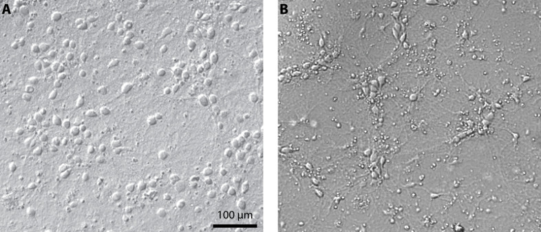
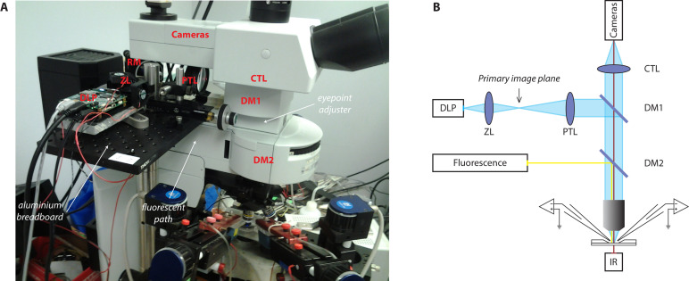
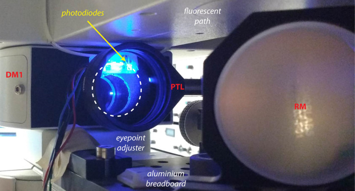
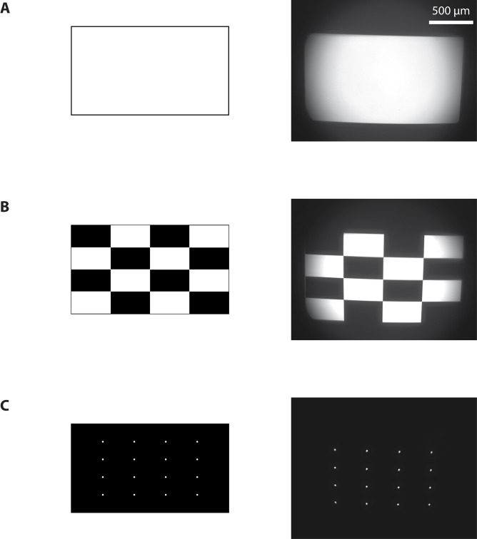
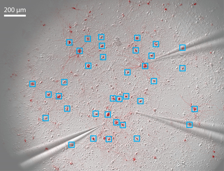
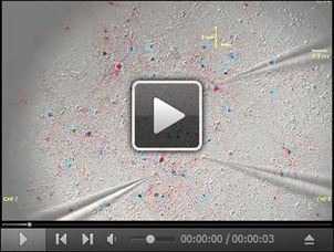
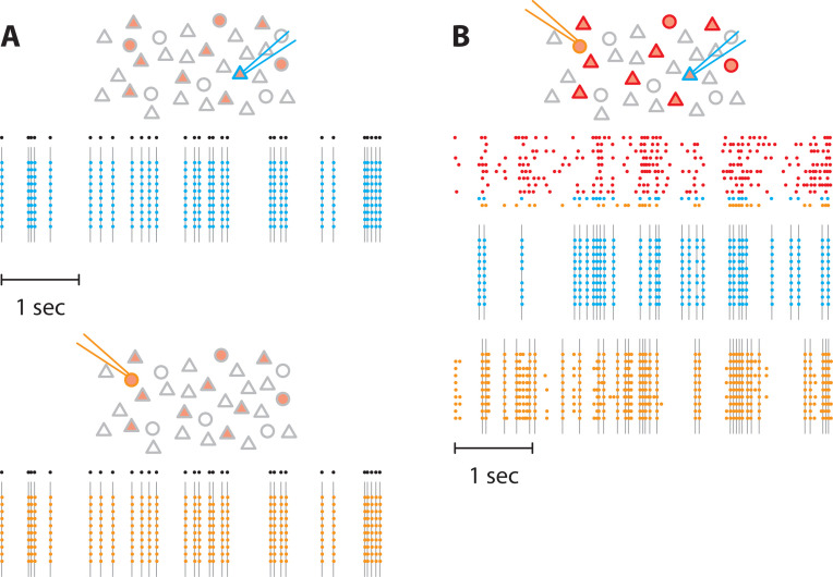
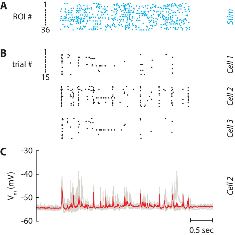
We studied a network of cortical neurons in culture and developed an innovative optical device to stimulate optogenetically a large neuronal population with both spatial and temporal precision. We first describe how to culture primary neurons expressing channelrhodopsin. We then detail the optogenetic setup based on the workings of a fast Digital Light Processing (DLP) projector. The setup is able to stimulate tens to hundreds neurons with independent trains of light pulses that evoked action potentials with high temporal resolution. During photostimulation, network activity was monitored using patch-clamp recordings of up to 4 neurons. The experiment is ideally suited to study recurrent network dynamics or biological processes such as plasticity or homeostasis in a network of neurons when a sub-population is activated by distinct stimuli whose characteristics (correlation, rate, and, size) were finely controlled.

Keywords: Optogenetics; Patterned optical stimulation; Primary culture of neurons.


Figures

Figure 1.
Figure 1.. Culture of primary cortical neurons at 14 days in vitro.
Representative examples of a healthy culture where cell bodies are well defined (A) and a culture where the coverslip coating was not achieved properly which resulted in cell aggregates and debris (B).
Figure 2.
Figure 2.. Photostimulation setup.
A. Picture of the photostimulation setup; B. Schematic of the light path. DLP, Digital Light Processing projector; ZL, zoom lens; PTL, projector tube lens; EPA, eyepoint adjuster; CTL, camera tube lens; DM, dichroic mirror; RM, reflective mirror.
Figure 3.
Figure 3.. Photodiodes system.
The photodiodes are placed next to the entry port of the microscope and collect a fraction of the beam coming from the projector (dashed line).
Figure 4.
Figure 4.. Calibration of the photostimulation setup.
Images sent to the projector (left) and resulting images from the camera (right) when a full-on (A), ANSI checkerboard (B), or a 4 x 4 white dots (C) pattern were projected.
Figure 5.
Figure 5.. Pattern stimulation of the neuronal culture.
36 regions of interest (ROIs) are drawn around stimulated neurons (blue squares). 3 patch-clamp electrodes record spikes and/or subthreshold membrane potential of non-stimulated neurons.
Video 1.
Video 1.. Pattern stimulation of the neuronal culture.
36 regions of interest (ROIs) are drawn around stimulated neurons (blue squares). Each stimulated neuron is activated by a train of light pulses (the actual stimulus is shown in blue on the top left corner). Three neurons are recorded simultaneously in cell-attached mode (yellow raster plots of evoked spikes in each corner, 15 repetitions of the stimulus) and then in whole-cell configuration (yellow trace of membrane potential, average of 7 repetitions).
Figure 6.
Figure 6.. Pattern stimulation efficacy in evoking spikes in ChR2-expressing neurons.
In the following schematics, neurons expressing ChR2 are shown in light red. Recorded neurons are designated by the recording pipets. Light-stimulated neurons have colored contours. A. A train of action potentials (black dots) was applied to two different neurons expressing ChR2 (blue and orange) independently (top and bottom, respectively). Several trials were realized to confirm the faithful stimulation of neurons of interest. Colored dots denote recorded action potential and grey lines the light flashes. No action potentials were observed in the non-stimulated neuron. B. Then, we stimulated 11 neurons in the network. We generated 11 spike trains (top) that were applied to neurons expressing ChR2, including the two previous neurons. Here, we used correlated spike trains for the stimulus (the average correlation between spike trains was C = 0.5). The blue and the orange neurons were recorded simultaneously.
Figure 7.
Figure 7.. Independent stimulation of the neuronal network.
A. Spike trains which are used for stimulating the 36 selected neurons in Figure 5. Each line is a train of light pulses applied to a given ROI. B. Raster plot of three neurons recorded simultaneously. Each line represents a given repetition of the same stimulus (15 repetitions). C. Corresponding membrane potential of Cell 2. Each repetition (7 repetitions) is drawn as a grey line and the average is displayed in red. The patch pipet contained the Na+ channel blocker QX-314 to block spikes and isolate postsynaptic potentials.

References

    1. Anselmi F., Banerjee A. and Albeanu D. F.(2015). Patterned photostimulation in the brain. In: Douglass, A. D.(Ed.). New Techniques in Systems Neuroscience. Springer; 235-270.
    1. Barral J. and Reyes A.(2016). Synaptic scaling rule preserves excitatory-inhibitory balance and salient neuronal network dynamics. Nat Neurosci 19(12): 1690-1696. - PubMed
    1. Boyden E. S., Zhang F., Bamberg E., Nagel G. and Deisseroth K.(2005). Millisecond-timescale, genetically targeted optical control of neural activity. Nat Neurosci 8(9): 1263-1268. - PubMed
    1. Brewer G. J., Torricelli J. R., Evege E. K. and Price P. J.(1993). Optimized survival of hippocampal neurons in B27-supplemented neurobasal, a new serum-free medium combination. J Neurosci Res 35(5): 567-576. - PubMed
    1. Farah N., Reutsky I. and Shoham S.(2007). Patterned optical activation of retinal ganglion cells. Conf Proc IEEE Eng Med Biol Soc 2007: 6368-6370. - PubMed