. 2017 Aug 10;4(4):ENEURO.0232-17.2017.
doi: 10.1523/ENEURO.0232-17.2017. eCollection 2017 Jul-Aug.

Super-Resolution Microscopy Reveals a Nanoscale Organization of Acetylcholine Receptors for Trans-Synaptic Alignment at Neuromuscular Synapses

Affiliations

Super-Resolution Microscopy Reveals a Nanoscale Organization of Acetylcholine Receptors for Trans-Synaptic Alignment at Neuromuscular Synapses

Amanda L York et al. eNeuro. .

Abstract

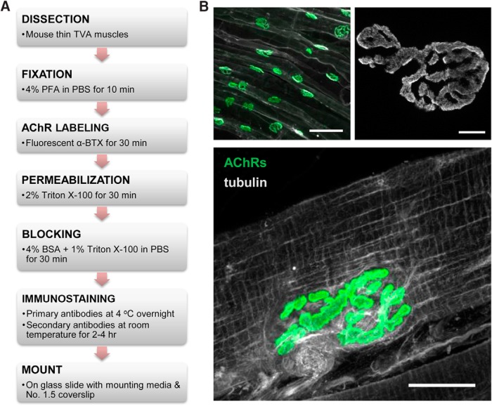
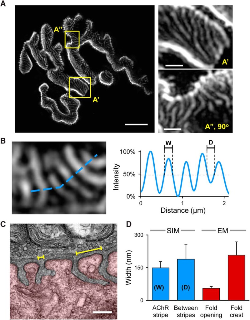
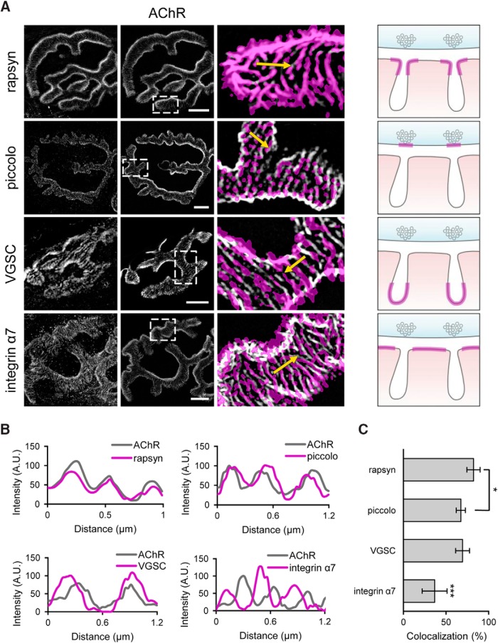
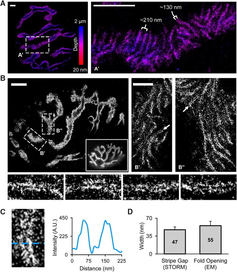
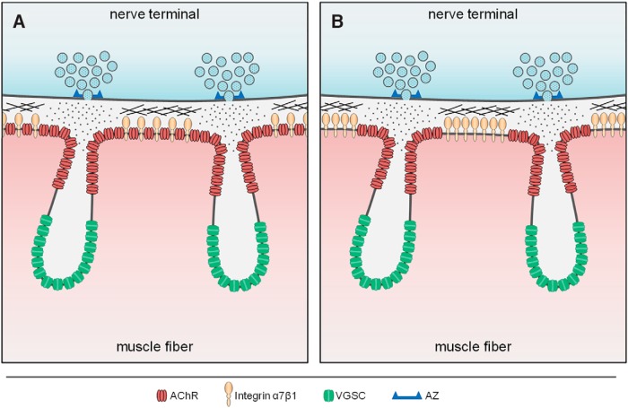
The neuromuscular junction (NMJ) is a chemical synapse formed between motoneurons and skeletal muscle fibers. The vertebrate NMJ uses acetylcholine (ACh) as the neurotransmitter and features numerous invaginations of the postsynaptic muscle membrane termed junctional folds. ACh receptors (AChRs) are believed to be concentrated on the crest of junctional folds but their spatial organization remains to be fully understood. In this study, we utilized super-resolution microscopy to examine the nanoscale organization of AChRs at NMJ. Using Structured Illumination Microscopy, we found that AChRs appear as stripes within the pretzel-shaped mouse NMJs, which however, do not correlate with the size of the crests of junctional folds. By comparing the localization of AChRs with several pre- and postsynaptic markers of distinct compartments of NMJs, we found that AChRs are not distributed evenly across the crest of junctional folds as previously thought. Instead, AChR stripes are more closely aligned with the openings of junctional folds as well as with the presynaptic active zone. Using Stochastic Optical Reconstruction Microscopy (STORM) for increased resolution, we found that each AChR stripe contains an AChR-poor slit at the center that is equivalent to the size of the opening of junctional folds. Together, these findings indicate that AChRs are largely localized to the edges of crests surrounding the opening of folds to align with the presynaptic active zones. Such a nanoscale organization of AChRs potentially enables trans-synaptic alignment for effective synaptic transmission of NMJs.

Keywords: NMJ; junctional folds; spatial distribution; super-resolution microscopy; synaptic receptors.


Figures

None
Figure 1. Whole-mount immunostaining of the TVA muscle for reliable detection of antigens at the NMJ. A, The flowchart depicting the protocol used for clean and reliable immunostaining of muscle fibers. B, Example images of the immunostaining. Top left, A low magnification image shows NMJ innervation patterns along the TVA muscles. Integrin α7 (white), used to highlight the membrane of individual muscle fibers, was colabeled with AChRs (green) to highlight the innervation pattern along the TVA muscle. Scale bar, 100 µm. Top right, A high magnification image of an individual NMJ shows the pretzel-shaped AChR distribution at the NMJ of a TVA muscle. Note that AChRs are not uniformly distributed. Scale bar, 5 µm. Bottom: AChRs (green) were colabeled with tubulin (white), an intracellular antigen, to show that the whole-mount method enables excellent antibody penetration to label the microtubule network inside the skeletal muscle. Scale bar, 20 µm.
Figure 2.
Figure 2.
AChRs are distributed in stripes that are not correlated with the crest of NMJ junctional folds. A, A representative 3D-SIM image showing the AChR-rich stripes separated by dark bands. Scale bar, 5 µm. The areas enclosed by yellow rectangles (A’, A”) are shown in a high magnification on the right. Scale bar, 1 µm. B, A small region of the AChR stripes (left panel) are used to generate the intensity profile shown on the right. The width of the AChR stripes (W) and the distance between two adjacent stripes (D) are measured and presented in the bar graph in D. C, A representative transmission electron micrograph of an NMJ from the TVA muscle. The junctional folds are clearly visible at the postsynaptic compartment (highlighted by red color). Numerous synaptic vesicles and mitochondria are present within the opposing presynaptic terminal. The average width of junctional fold openings and fold crests (yellow brackets) were manually quantified and presented in the bar graph in D. Scale bar, 0.2 µm. D, The bar graph summarizing the measurement results from the SIM data (blue bars, n = 4, >180 stripes) and EM data (red bars; n = 7, >40 folds). Error bars represent the SD.
Figure 3.
Figure 3.
Correlation of AChR distribution with specific pre- and postsynaptic markers at NMJs. A, B, Colocalization of AChRs (white) with various synaptic markers (magenta, threshold). Representative fluorescent images are shown on the right and the schematics on the right depict the known localization of each synaptic marker (magenta line) with respect to the junctional folds. Scale bars, 5 µm. The representative intensity profiles of AChRs (gray) and each of the synaptic markers (magenta) are shown in B. Four markers were examined: Rapsyn, an intracellular AChR scaffolding protein; piccolo, an active zone component; integrin α7, an integrin subunit involved in adhesion in NMJs. C, Quantification of the colocalization of each marker with AChRs. One-way ANOVA analysis: p = 2.22 × 10⁻⁵ (n = 4). Error bars represent the SD. Bonferroni analysis: *p = 0.012, ***p < 0.004.
Figure 4.
Figure 4.
Super-resolution imaging reveals AChRs are concentrated around the opening of junctional folds. A, 3D-STORM imaging of AChRs highlights the 3D nature of the postsynaptic membrane. The widths of AChR stripes (∼130 nm) and the distance between stripes (∼210 nm) is consistent with quantifications from our previous SIM data. A small region outlined by dashed rectangle (A’) is shown in a higher magnification on the right. Color scale bar indicates Z-depth. Scale bars, 2 µm. B, Representative STORM images of AChRs at a NMJ. Scale bar, 5 µm. Inset image shows the same NMJ imaged using widefield microscopy. Close-up regions (B’, B”) reveals a thin slit at the center of each AChR stripe (arrow). Scale bar, 1 µm. Bottom, close-up view of individual AChR stripes. C, Representative profile linescan further highlighting the slit at the center of each AChR stripe. D, Quantification of the width of the AChR stripe gap (measured from STORM data, n = 23) and the width of the junctional fold opening (measured from our TEM data, n = 50). Error bars represent the SD. Average width values at center of each bar.
Figure 5.
Figure 5.
Schematics showing the current view (A) and the proposed revision (B) of AChR distribution along junctional fold crests. Classically, it is believed that AChRs (red) are distributed across the entire junctional fold crest and partially down the sides of the infolded membrane and excluded from the trough of junctional folds where VGSC (green) are localized (A). However, we propose that AChRs are instead spatially restricted to the area immediately surrounding the opening of junctional folds and are segregated from the adhesion molecule integrin α7β1 (tan) located at the center-most part of fold crests (B). The spatial segregation of AChRs and integrin α7β1 could be beneficial to maintaining strong synaptic adhesion between the pre- and postsynaptic terminals. Furthermore, this subsynaptic organization would position AChRs directly opposite that of the active zone (AZ, blue bracket), and thus, in the prime position to receive and respond to acetylcholine (dots) release.

References

    1. Antolik C, Catino DH, O'Neill AM, Resneck WG, Ursitti JA, Bloch RJ (2007) The actin binding domain of ACF7 binds directly to the tetratricopeptide repeat domains of rapsyn. Neuroscience 145:56–65. 10.1016/j.neuroscience.2006.11.047 - DOI - PMC - PubMed
    1. Barik A, Lu Y, Sathyamurthy A, Bowman A, Shen C, Li L, Xiong WC, Mei L (2014) LRP4 is critical for neuromuscular junction maintenance. J Neurosci 34:13892–13905. 10.1523/JNEUROSCI.1733-14.2014 - DOI - PMC - PubMed
    1. Burden SJ, Sargent PB, McMahan UJ (1979) Acetylcholine receptors in regenerating muscle accumulate at original synaptic sites in the absence of the nerve. J Cell Biol 82:412–425. - PMC - PubMed
    1. Chen J, Mizushige T, Nishimune H (2012) Active zone density is conserved during synaptic growth but impaired in aged mice. J Comp Neur 520:434–452. 10.1002/cne.22764 - DOI - PMC - PubMed
    1. Couteaux R, Pecot-Dechavassine M (1970) [Synaptic vesicles and pouches at the level of “active zones” of the neuromuscular junction]. C R Acad Sci Hebd Seances Acad Sci D 271:2346–2349. - PubMed

Publication types

MeSH terms

LinkOut - more resources