. 2017 Dec;66(12):1563-1575.
doi: 10.1007/s00262-017-2050-7. Epub 2017 Aug 10.

Mucosa-associated invariant T cells infiltrate hepatic metastases in patients with colorectal carcinoma but are rendered dysfunctional within and adjacent to tumor microenvironment

Affiliations

Mucosa-associated invariant T cells infiltrate hepatic metastases in patients with colorectal carcinoma but are rendered dysfunctional within and adjacent to tumor microenvironment

Christopher R Shaler et al. Cancer Immunol Immunother. 2017 Dec.

Abstract

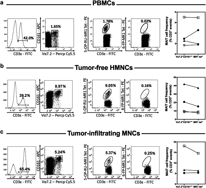
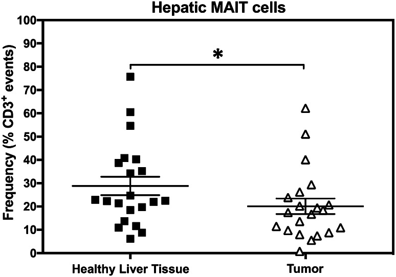
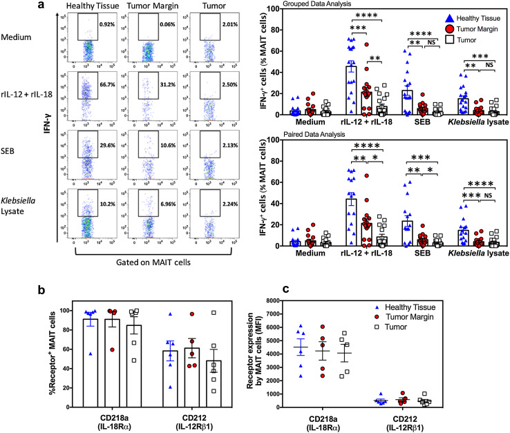
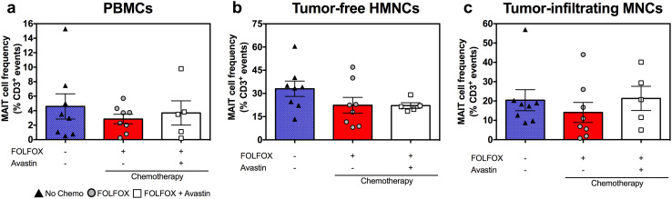
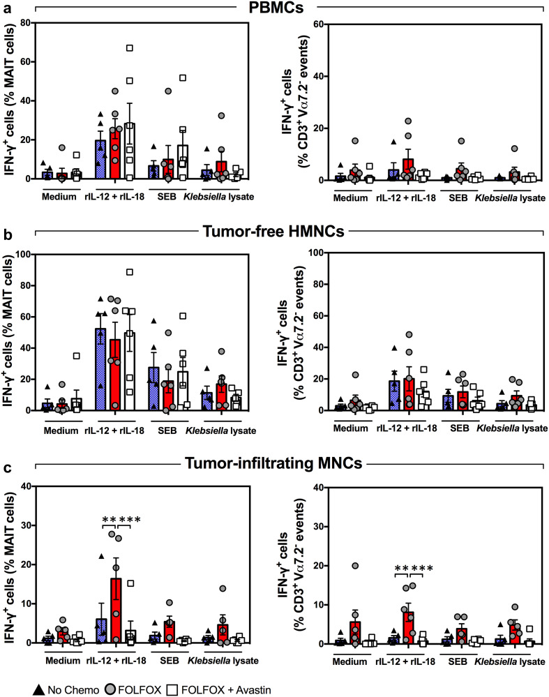
Mucosa-associated invariant T (MAIT) cells are innate-like T lymphocytes that are unusually abundant in the human liver, a common site of colorectal carcinoma (CRC) metastasis. However, whether they contribute to immune surveillance against colorectal liver metastasis (CRLM) is essentially unexplored. In addition, whether MAIT cell functions can be impacted by chemotherapy is unclear. These are important questions given MAIT cells' potent immunomodulatory and inflammatory properties. Herein, we examined the frequencies and functions of peripheral blood, healthy liver tissue, tumor-margin and tumor-infiltrating MAIT cells in 21 CRLM patients who received no chemotherapy, FOLFOX, or a combination of FOLFOX and Avastin before they underwent liver resection. We found that MAIT cells, defined as CD3ε+Vα7.2+CD161++ or CD3ε+MR1 tetramer+ cells, were present within both healthy and tumor-afflicted hepatic tissues. Paired and grouped analyses of samples revealed the physical proximity of MAIT cells to metastatic lesions to drastically influence their functional competence. Accordingly, unlike those residing in the healthy liver compartment, tumor-infiltrating MAIT cells failed to produce IFN-γ in response to a panel of TCR and cytokine receptor ligands, and tumor-margin MAIT cells were only partially active. Furthermore, chemotherapy did not account for intratumoral MAIT cell insufficiencies. Our findings demonstrate for the first time that CRLM-penetrating MAIT cells exhibit wide-ranging functional impairments, which are dictated by their physical location but not by preoperative chemotherapy. Therefore, we propose that MAIT cells may provide an attractive therapeutic target in CRC and that their ligands may be combined with chemotherapeutic agents to treat CRLM.

Keywords: Chemotherapy; Colon cancer; Immune surveillance; Liver metastasis; MAIT cells; Tumor-infiltrating lymphocytes.


Conflict of interest statement

The authors declare that they have no conflict of interest.

Figures

Fig. 1
Fig. 1
Peripheral blood and hepatic MAIT cells can be readily detected in CRC patients with liver metastasis. The frequency of MAIT cells was determined by flow cytometry among PBMCs (a), tumor-free, non-parenchymal hepatic mononuclear cells (b) and hepatic tumor-infiltrating mononuclear cells (c) isolated from patients with CRLM. MAIT cells were defined as CD3ε+Vα7.2+CD161++ or CD3ε+5-OP-RU-MR1 tetramer+ cells as indicated. 6-FP-MR1 tetramer was used as a staining control. Representative dot plots for each sample type are depicted (left panels). MAIT cell frequencies were calculated using both staining strategies for 3–4 patients (right panels). Values for matched samples (scatter plots with each patient being represented by a distinct symbol) are shown for comparison
Fig. 2
Fig. 2
MAIT cell frequencies are diminished within liver metastases of CRC. Tumor-free and tumor-infiltrating non-parenchymal mononuclear cells were isolated from the liver of each CRLM patient. MAIT cells were then enumerated for each patient’s sample set, and the Wilcoxon matched-pairs signed rank test was used to analyze data statistically. Error bars represent standard error of the mean (SEM)
Fig. 3
Fig. 3
The physical location of MAIT cells within CRLM-afflicted livers dictates their functional capacity. Non-parenchymal mononuclear cells harvested from healthy liver tissue, tumor margin and metastatic tumors of CRLM patients were left untreated or exposed to rIL-12+rIL-18, SEB or Klebsiella lysate. Twenty-four hours later, the frequencies of IFN-γ+ MAIT cells were determined by flow cytometry. Representative dot plots are illustrated, and statistical analyses of grouped and paired data sets were performed using two-way ANOVA and paired t tests, respectively (a). Baseline frequencies of CD212+ and CD218a+ hepatic MAIT cells (b) and the MFI of CD212 and CD218a staining (c) are also shown
Fig. 4
Fig. 4
Preoperative chemotherapy with FOLFOX or with FOLFOX plus Avastin does not lower blood and hepatic MAIT cell frequencies. PBMCs (a), tumor-free, non-parenchymal hepatic mononuclear cells (b) and hepatic tumor-infiltrating mononuclear cells (c) were isolated from CRLM patients who had received no chemotherapy (filled triangles), FOLFOX (filled circles) or FOLFOX plus Avastin (open squares) before they underwent liver resection surgery. The percentage of MAIT cells in each cell population was determined by flow cytometry. Error bars represent SEM
Fig. 5
Fig. 5
Baseline and inducible IFN-γ production by blood and hepatic MAIT cells is refractory to treatment with FOLFOX ± Avastin. PBMCs (a), tumor-free hepatic mononuclear cells (b) and tumor-infiltrating mononuclear cells (c) were isolated from CRLM patients who had received no chemotherapy (filled triangles), preoperative FOLFOX (filled circles) or FOLFOX plus Avastin (open squares). The ability of MAIT cells (left panels) and CD3+Vα7.2 conventional T cells (right panels) to produce IFN-γ in response to rIL-12+rIL-18, SEB or Klebsiella lysate was evaluated by flow cytometry. Error bars represent SEM

Similar articles

Cited by

References

    1. Siegel RL, Miller KD, Jemal A. Cancer statistics, 2016. CA Cancer J Clin. 2016;66(1):7–30. doi: 10.3322/caac.21332. - DOI - PubMed
    1. El-Shami K, Oeffinger KC, Erb NL, Willis A, Bretsch JK, Pratt-Chapman ML, Cannady RS, Wong SL, Rose J, Barbour AL, Stein KD, Sharpe KB, Brooks DD, Cowens-Alvarado RL. American Cancer Society colorectal cancer survivorship care guidelines. CA Cancer J Clin. 2015;65(6):428–455. doi: 10.3322/caac.21286. - DOI - PMC - PubMed
    1. Manfredi S, Lepage C, Hatem C, Coatmeur O, Faivre J, Bouvier AM. Epidemiology and management of liver metastases from colorectal cancer. Ann Surg. 2006;244(2):254–259. doi: 10.1097/01.sla.0000217629.94941.cf. - DOI - PMC - PubMed
    1. Misiakos EP, Karidis NP, Kouraklis G. Current treatment for colorectal liver metastases. World J Gastroenterol. 2011;17(36):4067–4075. doi: 10.3748/wjg.v17.i36.4067. - DOI - PMC - PubMed
    1. Sanchez-Castanon M, Er TK, Bujanda L, Herreros-Villanueva M. Immunotherapy in colorectal cancer: what have we learned so far? Clin Chim Acta. 2016;460:78–87. doi: 10.1016/j.cca.2016.06.027. - DOI - PubMed