. 2017 Aug 11;22(8):1323.
doi: 10.3390/molecules22081323.

Phosphate-Linked Silibinin Dimers (PLSd): New Promising Modified Metabolites

Affiliations

Phosphate-Linked Silibinin Dimers (PLSd): New Promising Modified Metabolites

Valeria Romanucci et al. Molecules. .

Abstract

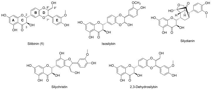
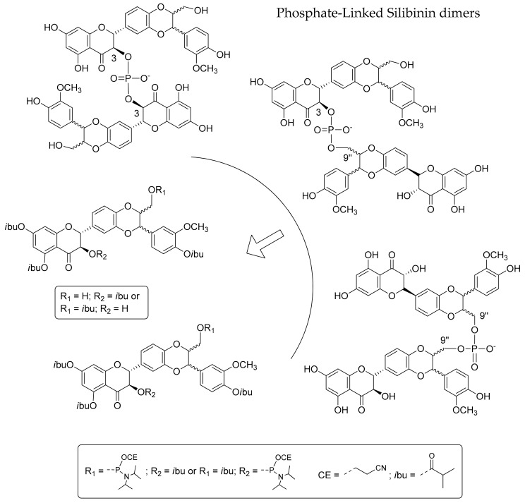
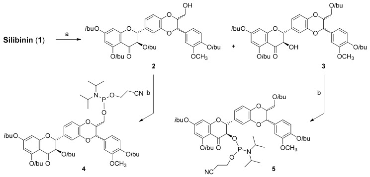
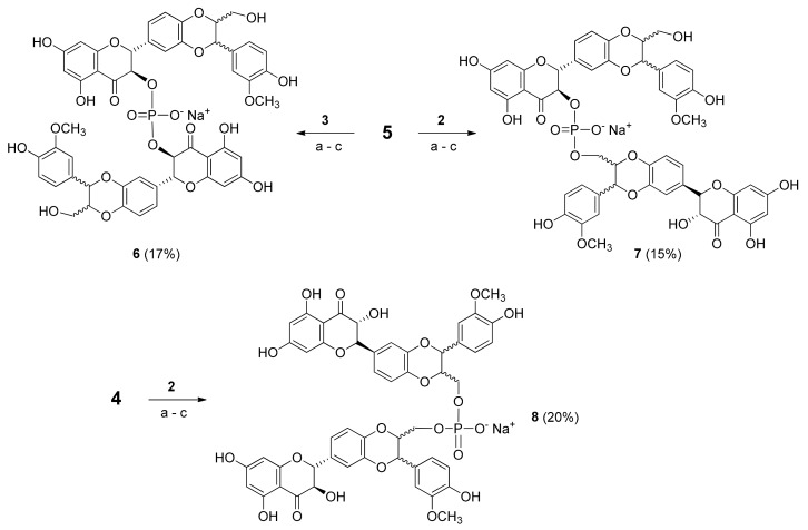
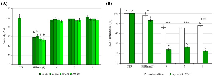
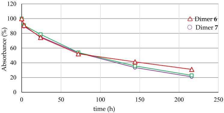
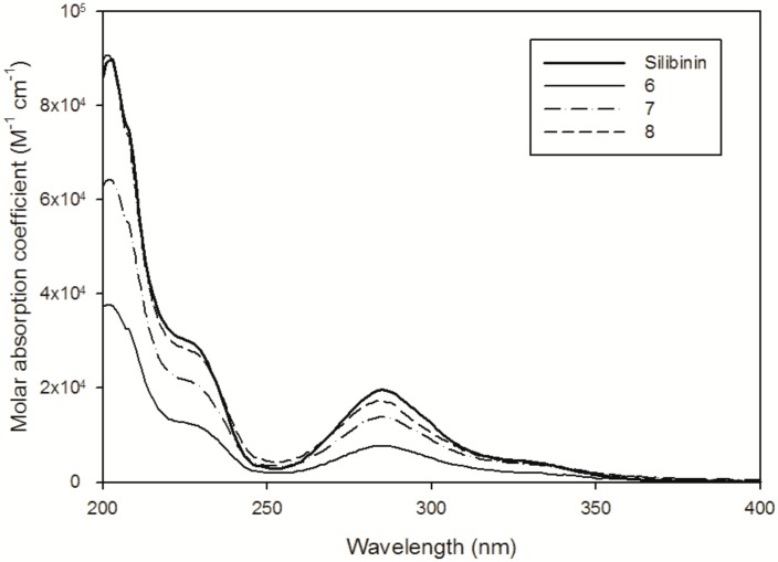
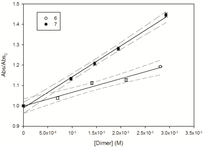
By exploiting the regioselective protection of the hydroxyl groups of silibinin along with the well-known phosphoramidite chemistry, we have developed an efficient strategy for the synthesis of new silibinin-modified species, which we have named Phosphate-Linked Silibinin Dimers (PLSd), in which the monomer units are linked by phosphodiester bonds. The antioxidant abilities of the new PLSd were estimated on HepG2 cells using DPPH free radical scavenging and xanthine/xanthine oxidase assays. The new phosphate-metabolites showed a higher anti-oxidant activity than the silibinin, as well as very low toxicity. The ability to scavenge reactive oxygen species (ROS) such as singlet oxygen () and hydroxyl radical () reveals that the two dimers are able to scavenge about two times more effectively than silibinin. Finally, solubility studies have shown that the PLSd present good water solubility (more than 20 mg·L-1) under circumneutral pH values, whereas the silibinin was found to be very poorly soluble (less than 0.4 mg·L-1) and not stable under alkaline conditions. Together, the above promising results warrant further investigation of the future potential of the PLSd as anti-oxidant metabolites within the large synthetic polyphenols field.

Keywords: oligoflavonoids; phosphodiester; radical scavengers; reactive oxygen species (ROS); silibinin; xanthine/xanthine oxidase assay.


Conflict of interest statement

The authors have no conflict of interest to declare.

Figures

Figure 1
Figure 1
Structures of all components of silymarin.
Scheme 1
Scheme 1
Retrosynthetic scheme of Phosphate-Linked Silibinin dimers.
Scheme 2
Scheme 2
Synthesis of silibinin building blocks. Reagents and conditions: (a) isobutyryl chloride, DIEA, THF 0 °C; (b) 2-cyanoethyl-N,N-diisopropylaminochlorophosphoramidite, DIEA, DCM, rt.
Scheme 3
Scheme 3
Synthesis of Phosphate-Linked Silibinin dimers 68, Reagents and conditions: (a) 0.45 M DCI, DCM, rt (b) 5.5 M TBHP in decane, rt (c) 28% NH4OH aqueous solution and CH3OH (1:1, v:v), 50 °C.
Figure 2
Figure 2
(A) The effects of 24 h treatment on HepG2 cell viability as determined by MTT assay. Different letters indicate significant differences among control and different concentrations (One way ANOVA, p < 0.05); (B) Protection against XO-induced intracellular ROS generation: DCF fluorescence was measured in basal conditions (white bars) or following cells exposure to X/XO system for 3 h (green bars). The cells were pretreated with each dimer (68) at concentrations of 20 μM. The values are expressed as % compared to the fluorescence value observed in control untreated cells which was set at 100. Asterisk indicates significant differences between basal conditions and exposure to X/XO, *: p < 0.05, ***: p < 0.001; different letters indicate significant differences among control, silibinin (1), 6, 7 and 8 within basal conditions and exposure to X/XO (Two way ANOVA).
Figure 3
Figure 3
Abundance (%) of the dimers 68 treated with human serum at 37 °C and analysed by RP-HPLC (see Section 3.13).
Figure 4
Figure 4
UV-Vis spectra of the silibinin in H2O-CH3CN (98:2) solution and the dimers in H2O at pH 6.5.
Figure 5
Figure 5
Abs/Abs0 measured at 475 nm as a function of 6 (PLSd 3-3) and 7 (PLSd 3-9′′). The solid line represents the linear fit of the experimental data, and dashed lines denote the 95% confidence interval of this fit.
Figure 6
Figure 6
Emission spectrum reaching the solution (1000 W xenon lamp equipped with a monochromator) and absorption spectrum of a 10 µM RB solution.

References

    1. Quideau S., Deffieux D., Douat-Casassus C., Pouységu L. Plant polyphenols: Chemical properties, biological activities, and synthesis. Angew. Chem. Int. Ed. 2011;50:586–621. doi: 10.1002/anie.201000044. - DOI - PubMed
    1. Bernini R., Fernanda C., Barontini M., Tofani D., Balducci V., Gambacorta A. Synthesis and structure/antioxidant activity relationship of novel catecholic antioxidant structural analogues to hydroxytyrosol and its lipophilic esters. J. Agric. Food Chem. 2012;60:7408–7416. doi: 10.1021/jf301131a. - DOI - PubMed
    1. Bernini R., Merendino N., Romani A., Velotti F. Naturally occurring hydroxytyrosol: Synthesis and anticancer potential. Curr. Med. Chem. 2013;20:655–670. doi: 10.2174/092986713804999367. - DOI - PubMed
    1. Barontini M., Bernini R., Carastro I., Gentili P., Romani A. Synthesis and DPPH radical scavenging activity of novel compounds obtained from tyrosol and cinnamic acid derivatives. New J. Chem. 2014;38:809–816. doi: 10.1039/C3NJ01180A. - DOI
    1. Bernini R., Gilardini Montani M.S., Merendino N., Romani A., Velotti F. Hydroxytyrosol-derived compounds: A basis for the creation of new pharmacological agents for cancer prevention and therapy. J. Med. Chem. 2015;58:9089–9107. doi: 10.1021/acs.jmedchem.5b00669. - DOI - PubMed

MeSH terms

LinkOut - more resources