. 2017 Aug 11;18(8):1745.
doi: 10.3390/ijms18081745.

Human Dental Pulp Cells Differentiate toward Neuronal Cells and Promote Neuroregeneration in Adult Organotypic Hippocampal Slices In Vitro

Affiliations

Human Dental Pulp Cells Differentiate toward Neuronal Cells and Promote Neuroregeneration in Adult Organotypic Hippocampal Slices In Vitro

Li Xiao et al. Int J Mol Sci. .

Abstract

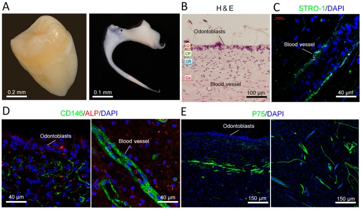
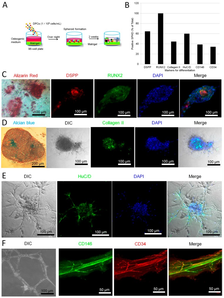
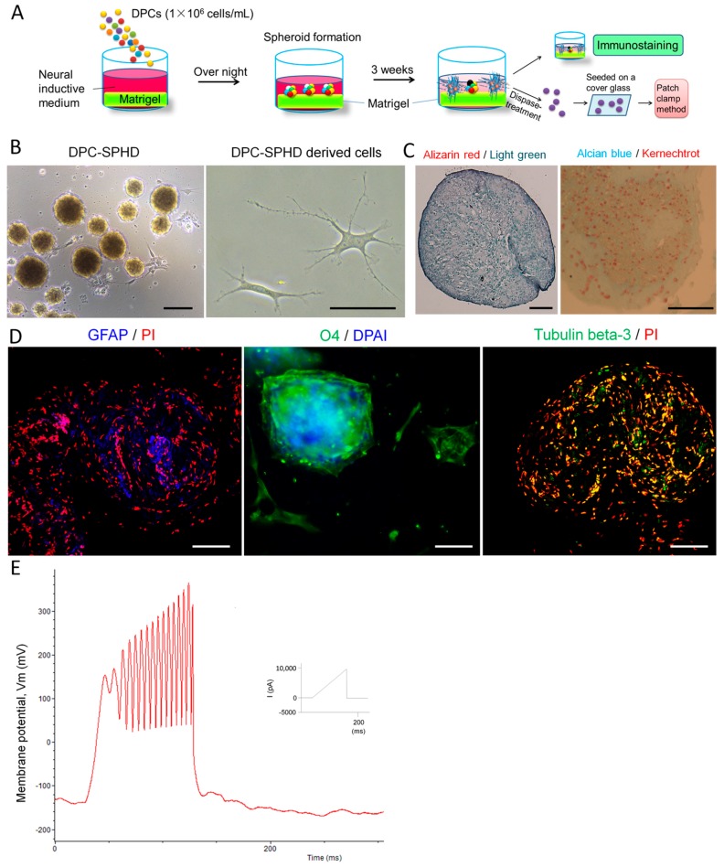
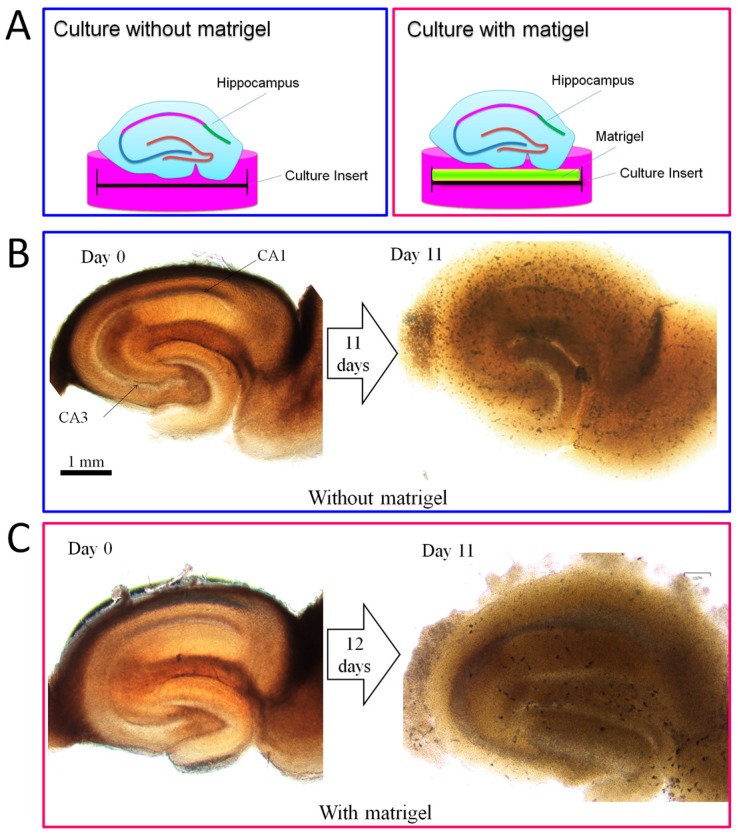
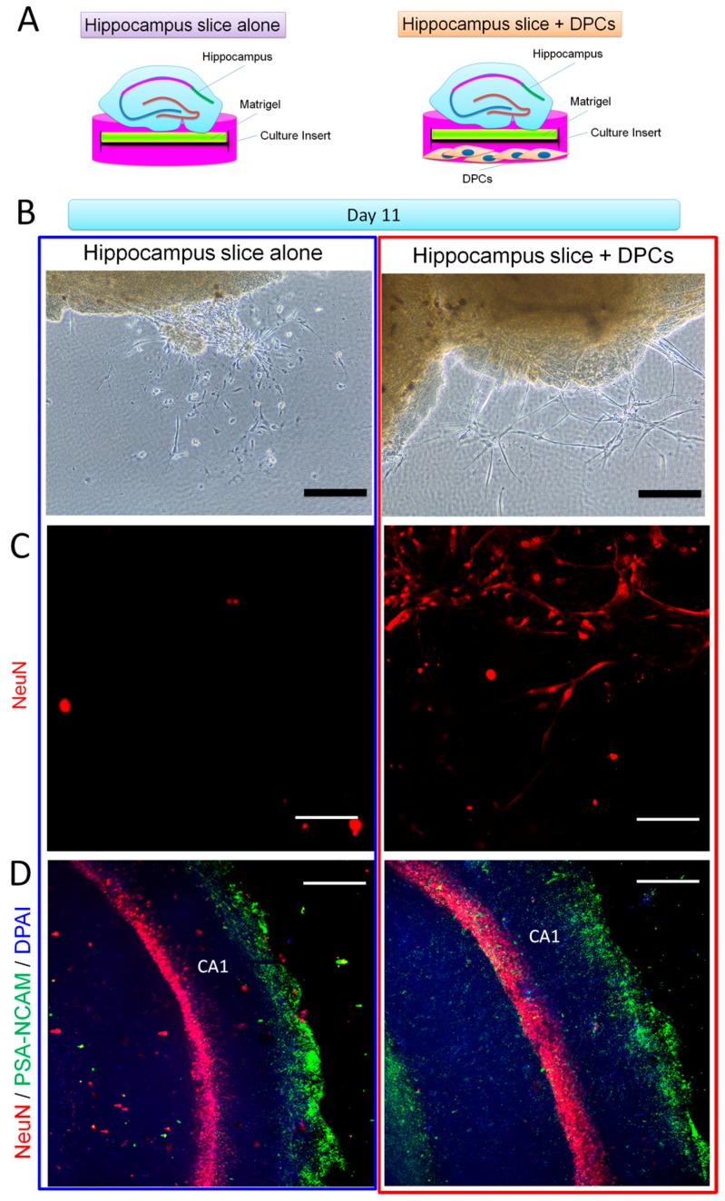
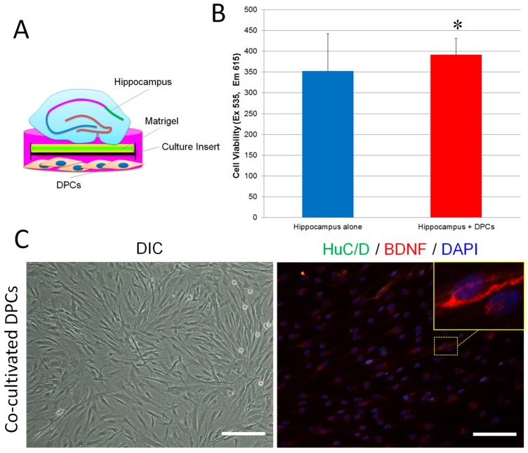
The adult mammalian central nerve system has fundamental difficulties regarding effective neuroregeneration. The aim of this study is to investigate whether human dental pulp cells (DPCs) can promote neuroregeneration by (i) being differentiated toward neuronal cells and/or (ii) stimulating local neurogenesis in the adult hippocampus. Using immunostaining, we demonstrated that adult human dental pulp contains multipotent DPCs, including STRO-1, CD146 and P75-positive stem cells. DPC-formed spheroids were able to differentiate into neuronal, vascular, osteogenic and cartilaginous lineages under osteogenic induction. However, under neuronal inductive conditions, cells in the DPC-formed spheroids differentiated toward neuronal rather than other lineages. Electrophysiological study showed that these cells consistently exhibit the capacity to produce action potentials, suggesting that they have a functional feature in neuronal cells. We further co-cultivated DPCs with adult mouse hippocampal slices on matrigel in vitro. Immunostaining and presto blue assay showed that DPCs were able to stimulate the growth of neuronal cells (especially neurons) in both the CA1 zone and the edges of the hippocampal slices. Brain-derived neurotrophic factor (BDNF), was expressed in co-cultivated DPCs. In conclusion, our data demonstrated that DPCs are well-suited to differentiate into the neuronal lineage. They are able to stimulate neurogenesis in the adult mouse hippocampus through neurotrophic support in vitro.

Keywords: adult hippocampal slice culture; co-cultivation; human dental pulp cells; neuronal regeneration.


Conflict of interest statement

The authors declare no conflict of interest.

Figures

Figure 1
Figure 1
Characterization of human dental pulp: stem cell distribution. (A) Digital photos of a human third molar tooth (left) and a human dental pulp from third molar (right); (B) Human dental pulp was stained with hematoxylin and eosin (H&E). O, odontoblastic zone; CP, cell poor zone; CR, cell rich zone; Co, core of the pulp; (C) Mesenchymal stem cell marker STRO-1 was expressed along the blood vessel. Nuclei were stained with DAPI (4′,6-diamidino-2-phenylindole); (D) Stem cell marker CD146 was expressed at the places near the odontoblasts and along the blood vessel. Alkaline phosphatase (ALP, marker for skeletal mineralization) was detected in the odontogenic zone; (E) Neuronal crest stem cell marker P75 was expressed in the core of dental pulp.
Figure 2
Figure 2
Multi-differentiation of dental pulp cell (DPC)-formed spheroids. Human dental pulp cells (1 × 10⁶) were seeded on matrigel in osteogenic medium overnight. The cells spontaneously formed spheroids. After further cultivation for 2 weeks, the spheroids differentiated into multi-lineage cell masses. (A) Experimental setup; (B) The spheroids were cultivated in osteogenic medium in 96-well plates for 14 days. The spheroids were followed by alizarin red, alcian blue and immunofluorescence staining. Five wells were prepared for each marker. The markers (antibodies) include DSPP, RUNX2, collagen II, HuC/D, CD146 and CD34. The positive cell masses for each marker were counted in every 10 spheroids per well × 5 wells/antibody; (CF) Typical images of differentiated spheroids (n = 5). Data represent three independent experiments.
Figure 3
Figure 3
Expression of neural markers for differentiation and electrophysiological properties in DPC-formed spheroids on matrigel. DPCs were seeded on matrigel in neural inductive medium overnight. The cells spontaneously formed spheroids in 96-well culture plates and were further cultivated for 3 weeks. (A) Experimental setup; (B) The spheroids in culture for 3 days. Scale bar = 50 µm; (C) Three weeks after neuronal induction, the spheroids were stained with alizarin red (nuclei were stained with light green) and alcian blue (nuclei were stained with kernechtrot). The spheroids were not stained by both alizarin red and alcian blue. Scale bar = 10 µm; (D) Expression of neural markers for differentiation in the spheroids after 3 weeks cultivation (immunofluorescence staining). Images were taken by LSM. The spheroids expressed the glia cell marker, GFAP, oligodendrocyte, O4, and the neuron marker, tubulin β-3. Nuclei were stained with propidium iodide (PI) or DAPI. Scale bar = 25 µm. DPC-SPHD, DPC-formed spheroids; (E) Electrophysiological characteristics of DPC-formed spheroids. Patch-clamp recordings of spheroid-derived cells. After being cultivated in neural inductive media for 3 weeks, spheroids were decomposed into single cells by dispase-treatment. The cells were seeded on a cover glass overnight to adhere to the glass surface. Action potential properties were analyzed by whole-cell patch-clamp recordings. Membrane potential oscillations were generated by ramp protocol using the current clamp mode with 10 nA maximal amplitude in spheroid-derived cells.
Figure 4
Figure 4
A novel method for long-term culture of adult mouse hippocampal slices in vitro. (A) Experimental setup; (B) Adult mouse hippocampal slices were cultivated on the culture insert without matrigel. After 11 days of cultivation, the CA1 and CA3 zones had disappeared; (C) Adult mouse hippocampal slices were cultivated on a matrigel-coated culture insert. After 11 days of cultivation, the CA1 and CA3 zones were still clear.
Figure 5
Figure 5
Effect of DPCs on neurogenesis in adult mouse hippocampal slices in vitro. (A) Experimental setup. Mouse hippocampus slices were cultivated on matrigel with DPCs or by themselves for 11 days, followed by immunofluorescence staining; (B) Images were taken by a phase contrast microscope. Hippocampus slice-derived cells showed glia cell-like morphology when cultivated alone (left). However, when the hippocampus slice co-cultivated with DPCs, the derived cells exhibited neuron-like morphology and formed networks. Scale bar = 50 µm (C) Immunofluorescence staining showed that cells derived from the co-cultivated hippocampus slice are positive to the mature neuron mark NeuN (right). However, cells derived from the single cultivated hippocampus did not react with NeuN (left). Images were taken by LSM. Scale bar = 25 µm; (D) In solo or co-cultivated hippocampus slices, cells reacted with NeuN and PSA-NCAM (a marker of developing and migrating neurons) antibodies. Images were taken by LSM. Scale bar = 50 µm.
Figure 6
Figure 6
DPCs promoted cell growth in adult mouse hippocampal slices in vitro and expressed BDNF. (A) Experimental setup; (B) Adult hippocampal slices were cultivated on matrigel-coated culture inserts (5 slices/well) with or without DPCs for 17 days. The culture inserts were transferred into new 6-well plates and followed by presto blue assay. Data are presented as mean ± SD. * p < 0.05; (C) DPCs kept fibroblast morphology while co-cultivated with hippocampal slices (left, magnification = 20×). Immunofluorescence staining showed that some DPCs were positive to anti-BDNF antibodies. The expression of HuC/D was hardly detected in co-cultivated DPCs (right, magnification = 20×). Scale bar = 50 µm

References

    1. Horner P.J., Gage F.H. Regenerating the damaged central nervous system. Nature. 2000;407:963–970. - PubMed
    1. Tanaka E.M., Ferretti P. Considering the evolution of regeneration in the central nervous system. Nat. Rev. Neurosci. 2009;10:713–723. doi: 10.1038/nrn2707. - DOI - PubMed
    1. Enciu A.M., Nicolescu M.I., Manole C.G., Mureşanu D.F., Popescu L.M., Popescu B.O. Neuroregeneration in neurodegenerative disorders. BMC Neurol. 2011;11:75. doi: 10.1186/1471-2377-11-75. - DOI - PMC - PubMed
    1. Xiao L., Saiki C., Ide R. Stem cell therapy for central nerve system injuries: Glial cells hold the key. Neural. Regen. Res. 2014;9:1253–1260. doi: 10.4103/1673-5374.137570. - DOI - PMC - PubMed
    1. Tam R.Y., Fuehrmann T., Mitrousis N., Shoichet M.S. Regenerative therapies for central nervous system diseases: A biomaterials approach. Neuropsychopharmacology. 2014;39:169–188. doi: 10.1038/npp.2013.237. - DOI - PMC - PubMed

MeSH terms