

Molecular basis for the functions of a bacterial MutS2 in DNA repair and recombination
- PMID: 28800560
- PMCID: PMC5787856
- DOI: 10.1016/j.dnarep.2017.07.004
Molecular basis for the functions of a bacterial MutS2 in DNA repair and recombination
Abstract
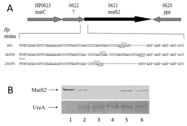
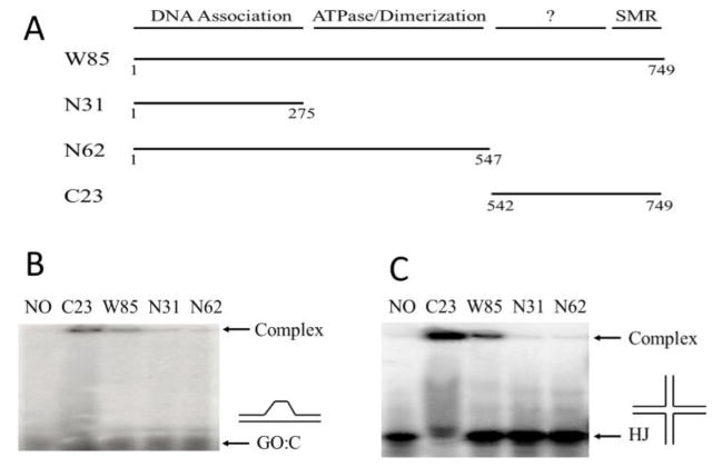
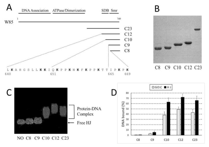
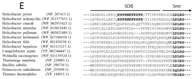
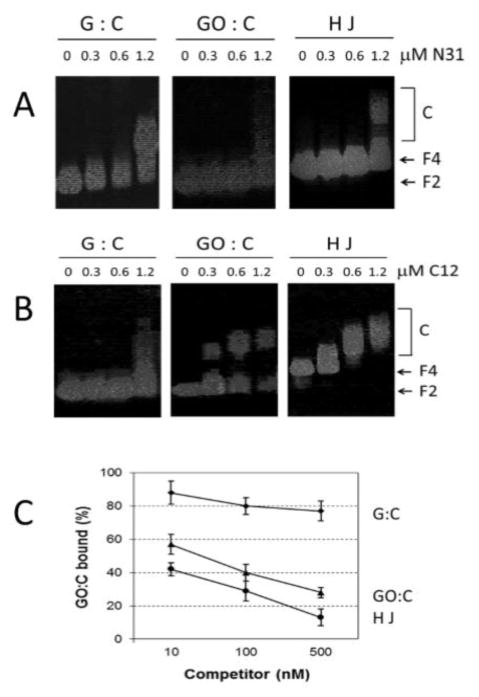
Bacterial MutS2 proteins, consisting of functional domains for ATPase, DNA-binding, and nuclease activities, play roles in DNA recombination and repair. Here we observe a mechanism for generating MutS2 expression diversity in the human pathogen Helicobacter pylori, and identify a unique MutS2 domain responsible for specific DNA-binding. H. pylori strains differ in mutS2 expression due to variations in the DNA upstream sequence containing short sequence repeats. Based on Western blots, mutS2 in some strains appears to be co-translated with the upstream gene, but in other strains (e.g. UA948) such translational coupling does not occur. Accordingly, strain UA948 had phenotypes similar to its ΔmutS2 derivative, whereas expression of MutS2 at a separate locus in UA948 (the genetically complemented strain) displayed a lower mutation rate and lower transformation frequency than did ΔmutS2. A series of truncated HpMutS2 proteins were purified and tested for their specific abilities to bind 8-oxoG-containing DNA (GO:C) and Holiday Junction structures (HJ). The specific DNA binding domain was localized to an area adjacent to the Smr nuclease domain, and it encompasses 30-amino-acid-residues containing a "KPPKNKFKPPK" motif. Gel shift assays and competition assays supported that a truncated version of HpMutS2-C12 (∼12kDa protein containing the specific DNA-binding domain) has much greater capacity to bind to HJ or GO:C DNA than to normal double stranded DNA. By studying the in vivo roles of the separate domains of HpMutS2, we observed that the truncated versions were unable to complement the ΔmutS2 strain, suggesting the requirement for coordinated function of all the domains in vivo.
Keywords: DNA recombination; Helicobacter pylori; MutS2; Oxidative DNA damage; Specific DNA binding; Translational coupling.
Copyright © 2017 Elsevier B.V. All rights reserved.
Conflict of interest statement
The authors declare no conflict of interest.
Figures
Similar articles
-
The nuclease activities of both the Smr domain and an additional LDLK motif are required for an efficient anti-recombination function of Helicobacter pylori MutS2.Mol Microbiol. 2015 Jun;96(6):1240-56. doi: 10.1111/mmi.13003. Epub 2015 Apr 23. Mol Microbiol. 2015. PMID: 25800579
-
Mutations in the nucleotide binding and hydrolysis domains of Helicobacter pylori MutS2 lead to altered biochemical activities and inactivation of its in vivo function.BMC Microbiol. 2016 Feb 3;16:14. doi: 10.1186/s12866-016-0629-3. BMC Microbiol. 2016. PMID: 26843368 Free PMC article.
-
Suppression of homologous and homeologous recombination by the bacterial MutS2 protein.Mol Cell. 2005 Jan 7;17(1):113-20. doi: 10.1016/j.molcel.2004.11.035. Mol Cell. 2005. PMID: 15629722
-
Genetic battle between Helicobacter pylori and humans. The mechanism underlying homologous recombination in bacteria, which can infect human cells.Microbes Infect. 2014 Oct;16(10):833-9. doi: 10.1016/j.micinf.2014.08.001. Epub 2014 Aug 14. Microbes Infect. 2014. PMID: 25130723 Review.
-
DNA mismatch repair: molecular mechanisms and biological function.Annu Rev Microbiol. 2003;57:579-608. doi: 10.1146/annurev.micro.57.030502.090847. Annu Rev Microbiol. 2003. PMID: 14527292 Review.
Cited by
-
A bacterial DNA repair pathway specific to a natural antibiotic.Mol Microbiol. 2019 Feb;111(2):338-353. doi: 10.1111/mmi.14158. Epub 2018 Nov 28. Mol Microbiol. 2019. PMID: 30379365 Free PMC article.
-
Genome and Methylome analysis of a phylogenetic novel Campylobacter coli cluster with C. jejuni introgression.Microb Genom. 2021 Oct;7(10):000679. doi: 10.1099/mgen.0.000679. Microb Genom. 2021. PMID: 34661518 Free PMC article.
-
Putative MutS2 Homologs in Algae: More Goods in Shopping Bag?J Mol Evol. 2024 Dec;92(6):815-833. doi: 10.1007/s00239-024-10210-y. Epub 2024 Oct 4. J Mol Evol. 2024. PMID: 39365456
-
α-Difluoromethylornithine reduces gastric carcinogenesis by causing mutations in Helicobacter pylori cagY.Proc Natl Acad Sci U S A. 2019 Mar 12;116(11):5077-5085. doi: 10.1073/pnas.1814497116. Epub 2019 Feb 25. Proc Natl Acad Sci U S A. 2019. PMID: 30804204 Free PMC article.
-
Expansion of the MutS Gene Family in Plants.bioRxiv [Preprint]. 2024 Jul 20:2024.07.17.603841. doi: 10.1101/2024.07.17.603841. bioRxiv. 2024. Update in: Plant Cell. 2025 Jul 1;37(7):koae277. doi: 10.1093/plcell/koae277. PMID: 39071318 Free PMC article. Updated. Preprint.
References
-
- Suerbaum S. Genetic variability within Helicobacter pylori. International journal of medical microbiology: IJMM. 2000;290:175–181. - PubMed
-
- Chaturvedi R, Cheng Y, Asim M, Bussiere FI, Xu H, Gobert AP, Hacker A, Casero RA, Jr, Wilson KT. Induction of polyamine oxidase 1 by Helicobacter pylori causes macrophage apoptosis by hydrogen peroxide release and mitochondrial membrane depolarization. The Journal of biological chemistry. 2004;279:40161–40173. - PubMed
-
- Allen LA, Beecher BR, Lynch JT, Rohner OV, Wittine LM. Helicobacter pylori disrupts NADPH oxidase targeting in human neutrophils to induce extracellular superoxide release. J Immunol. 2005;174:3658–3667. - PubMed
MeSH terms
Substances
Grants and funding
LinkOut - more resources
Full Text Sources
Other Literature Sources
