. 2017 Aug 11;13(8):e1006560.
doi: 10.1371/journal.ppat.1006560. eCollection 2017 Aug.

Expression of the RNA-binding protein RBP10 promotes the bloodstream-form differentiation state in Trypanosoma brucei

Affiliations

Expression of the RNA-binding protein RBP10 promotes the bloodstream-form differentiation state in Trypanosoma brucei

Elisha Mugo et al. PLoS Pathog. .

Abstract

In nearly all eukaryotes, cellular differentiation is governed by changes in transcription, and stabilized by chromatin and DNA modification. Gene expression control in the pathogen Trypanosoma brucei, in contrast, relies almost exclusively on post-transcriptional mechanisms, so RNA binding proteins must assume the burden that is usually borne by transcription factors. T. brucei multiply in the blood of mammals as bloodstream forms, and in the midgut of Tsetse flies as procyclic forms. We show here that a single RNA-binding protein, RBP10, promotes the bloodstream-form trypanosome differentiation state. Depletion of RBP10 from bloodstream-form trypanosomes gives cells that can grow only as procyclic forms; conversely, expression of RBP10 in procyclic forms converts them to bloodstream forms. RBP10 binds to procyclic-specific mRNAs containing an UAUUUUUU motif, targeting them for translation repression and destruction. Products of RBP10 target mRNAs include not only the major procyclic surface protein and enzymes of energy metabolism, but also protein kinases and stage-specific RNA-binding proteins: this suggests that alterations in RBP10 trigger a regulatory cascade.


Conflict of interest statement

The authors have declared that no competing interests exist.

Figures

Fig 1
Fig 1. Tethering of the C-terminal portion of RBP10 to a reporter mRNA inhibits its translation and causes its destruction.
A. Various fragments of RBP10 were inducibly expressed with an N-terminal lambdaN peptide and a C-terminal myc tag, in a cell line expressing a CAT reporter mRNA with 5 boxB sequences in the 3'-UTR. Cartoons showing the fragments are on the left. The numbers above the full-length protein are amino acid residues. The effects on the amounts CAT mRNA and CAT protein were measured by Northern blot and enzyme assay, respectively. Levels are expressed as mean ± standard deviation for at least 3 replicates, relative to a line with no lambdaN peptide protein (top bars). B. Bloodstream-form trypanosomes expressing the reporter in (A), and with tetracycline-inducible expression of lamdaN-RBP10-F3, were used. The reporter with or without tethered protein is shown at the top. Lysates from cells grown without (left) or with (right) tetracycline were subjected to sucrose gradient centrifugation. The panel below the cartoons show the absorption profiles at 254 nm as the fractions were collected. The migration of CAT and beta-tubulin (TUB) mRNAs on the gradient was detected by Northern blotting; representative blots are shown. The graphs show Northern signals, expressed as the percentage of the total signal, with arithmetic mean and standard deviation for three independent biological replicates.
Fig 2
Fig 2. RBP10 targets a UAUUUUUU motif.
A) Abundances of mRNAs that were at least 3x enriched in both RBP10-bound fraction, relative to those that were less than 1x enriched. B) The 3'-UTRs of 188 mRNAs that were at least 3x enriched in both RBP10-bound samples (Supplementary S2 Table) were compared with those of mRNAs that did not bind (<0.7x enrichment in both experiments) using DREME. Only 3'-UTRs annotated in tritrypDB were used (S1 and S2 Text) and some were not mapped with sufficient confidence to be included. The best-scoring motif found is shown in the inset. The graph shows numbers of UA(U)6 motifs in 255 manually annotated bound 3'-UTRs (S2 Table). One mRNA each had 7, 8 and 9 motifs. The numbers of motifs shown here are sometimes higher than with automatically annotated 3'-UTRs from the genome database, because some of the automatic 3'-UTRs are truncated. C) Effect of the UA(U)6 motif on RBP10 binding: median ± 25th percentiles, with 95% confidence limits and outliers. Database 3'-UTRs were analyzed. Since these are often truncated, and are missing for 30% of genes, the numbers of motifs are under-estimated. Significant differences (Student t-test) are shown. D) Reporters used in (E) and (F) [42]. The red bars are UAUUUUUU elements, which are highlighted in the 26mer sequence. E) RBP10 binding to the EP 3'-UTR requires the 26mer. Cells were UV-irradiated, and RBP10 was pulled down with anti-RBP10 (upper panel, Western blot). RNAs were detected by reverse transcription and PCR using gene-specific primers (lower panel). If reverse transcriptase was omitted, no PCR band was obtained. F) rbp10 RNAi specifically affects expression of a reporter containing the 26mer. CAT activity was measured 17h and 24h after RNAi induction.
Fig 3
Fig 3. mRNAs bound to and/or regulated by RBP10.
Results for mRNA levels are displayed as proportional Venn diagrams made using http://www.eulerdiagrams.org/eulerAPE/. BS = Bloodstream form, PC = procyclic form. Categories were: Bound: S2 Table sheet 2. PC>BS and BS>PC: at least 2x significantly regulated in polysomal RNA. Values for increases after rbp10 RNAi in bloodstream forms (at least 2x significantly regulated), or RBP10-myc expression in procyclic forms (at least 1.5x significantly regulated), are from S3 Table. All data are from Lister 427 strain parasites. A) RBP10-bound mRNAs relative to those that are more abundant in procyclic forms and/or increase after rbp10 RNAi. B) Overlaps between mRNAs that are more abundant in bloodstream forms, and those that decreased after rbp10 RNAi in bloodstream forms or increased after RBP10 expression in procyclic forms. C) Overlaps between bound RNAs, procyclic-specific mRNAs, and the mRNAs that decreased after RBP10 expression in procyclic forms.
Fig 4
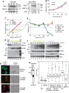
Fig 4. RBP10 depletion primes bloodstream forms for differentiation to procyclic forms.
A) Expression of PAD1 and RBP10 in trypanosomes incubated at maximal density in methyl cellulose-containing medium for 3 days (left panel), or after rbp10 RNAi (right panel). * = nonspecific band. B) Growth of trypanosomes incubated at maximal density in methyl cellulose-containing medium for 3 days, followed by cis-aconitate treatment for 17h at 27°C. Time 0 is the time of transfer to procyclic conditions. C) Cumulative growth curve of bloodstream-form (BS) trypanosomes with and without RNAi. WT = wild-type (tet repressor only); -RNAi: RNAi cell line with no tetracyline: +RNAi: RNAi cell line with tetracycline. D) Expression of RBP10, PIP39, and EP procyclin were measured by Western blotting up to 3d after tetracycline induction of RNAi. E) 17h after RNAi induction at 37°C in bloodstream form medium (+RNAi), or incubation at high density in procylic medium with 6mM cis-aconitate at 27°C (+CA), trypanosomes were placed in procyclic medium (MEM-pros) at 27°C with neither tetracycline nor cis-aconitate. The cell densities of the cultures were monitored. Negative controls were either wild-type or uninduced RNAi lines, without cis-aconitate. Cell densities are shown; they started to increase at 72h. F) Protein expression after transfer to procyclic medium at 27°C after 17h pre-treatment as in (E). G) Immunofluorescence: Cell morphology and expression of GPEET procyclin after 3d cultivation in procyclic medium. DNA is stained with DAPI. H) The cartoon shows the way in which the position of the kinetoplast, relative to the nucleus (P-K/K-N), was measured. The box plot shows the median with 25th percentiles and 95% confidence limits. Some outliers have been deleted. The full results, together with the numbers of trypanosomes measured, are in S8A Fig. BS—normal bloodstream forms; PC- normal procyclic forms; CA - 17h cis-aconitate pretreatment; RNAi or Ri; 17h rbp10 RNAi induction; +RBP10—induced expression of RBP10 for 2 days. There were no significant differences between the 3-day and 6-day differentiating populations. All other pairs were significantly different (Student t-test, p<0.05).
Fig 5
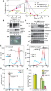
Fig 5. RBP10 expression in procyclic forms causes differentiation to bloodstream forms.
A) Cell counts in a typical experiment. Expression of RBP10-myc was induced using tetracycline at 27°C in procyclic-form medium. The left-hand panel is a cumulative growth curve. After 48h cells were transferred to bloodstream-form medium at 37°C; in the right-hand panel cell densities are shown. B) Expression of EP procyclin and RBP10 after induction of RBP10-myc expression. Cells were cultured in procyclic medium at 27°C. Expression of EP procyclin protein decreased after 48h of RBP10-myc induction, while untagged RBP10 becomes detectable. C) As in (B) except that culture continued for 3 days. Proteins that are expressed more in bloodstream forms than in procyclic forms were detected by Western blotting. TAO is trypanosome alternative oxidase; HNRNPH is an RNA-binding protein. VSG mRNA was detected by RT-PCR using a spliced leader primer and a primer that hybridizes to a conserved region within the VSG 3'-UTR. D) Trypanosomes were taken after 48h RBP10-myc induction. They were stained for EP procyclin and phosphorylated GPEET procyclin (green), VSG117 (magenta) and DNA (cyan). The stain is overlaid with differential interference contrast (grey). The panel shows a typical green procyclic form, a bloodstream form with two kinetoplasts, and a third cell which resembles a bloodstream form (terminal kinetoplast) but was not stained by anti-VSG117. This cell may express a VSG that does not react with the anti-VSG117 antibodies. E) Quantitation of surface EP procyclin by flow cytometry, comparing cells with (day 2) and without (day 0) induced RBP10-myc expression. F) Quantitation of surface phospho-GPEET procyclin by flow cytometry, comparing cells with (day 2) and without (day 0) induced RBP10-myc expression. G) Quantitation of staining with anti-VSG117 by flow cytometry, comparing cells with (day 2) and without (day 0) induced RBP10-myc expression. H) Measurement of surface protein expression by flow cytometry. Values for the different windows are mean and standard deviation from 4 replicates. Similar results were obtained by manual counting of stained smears (S8 Fig).
Fig 6
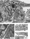
Fig 6. VSG expression after RBP10 induction in procyclic forms: Electron microscopy.
A) A sample field from procyclic-form cultures after 48h induced expression of RBP10. The thin arrows point to VSG coats and the thick ones to procyclic surfaces. B) Close-up of the boxed area in (A). C) Typical field showing the surface of a procyclic form. D) Typical field showing the surfaces of bloodstream forms.
Fig 7
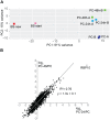
Fig 7. Analysis of transcriptomes of EATRO1125 with forced RBP10 expression.
A) Principal component analysis showing the clustering of procyclic forms, bloodstream forms and cells with induced RBP10 expression. B) Correlation between the effects of 24h and 48h RBP10 expression for 7000 unique genes. For each gene, the log2 of the ratio between the induced and uninduced cultures is shown.

References

    1. Simarro P, Cecchi G, Franco J, Paone M, Diarra A, Ruiz-Postigo J, et al. Estimating and Mapping the Population at Risk of Sleeping Sickness. PLoS Negl Trop Dis. 2012;6:e1859 doi: 10.1371/journal.pntd.0001859 - DOI - PMC - PubMed
    1. Shaw AP, Cecchi G, Wint GR, Mattioli RC, Robinson TP. Mapping the economic benefits to livestock keepers from intervening against bovine trypanosomosis in Eastern Africa. Prev Vet Med. 2014;113(2):197–210. Epub 2013/11/28. doi: 10.1016/j.prevetmed.2013.10.024 . - DOI - PubMed
    1. Dobson RJ, Dargantes AP, Mercado RT, Reid SA. Models for Trypanosoma evansi (surra), its control and economic impact on small-hold livestock owners in the Philippines. Int J Parasitol. 2009;39(10):1115–23. Epub 2009/03/11. doi: 10.1016/j.ijpara.2009.02.013 . - DOI - PubMed
    1. Fikru R, Andualem Y, Getachew T, Menten J, Hasker E, Merga B, et al. Trypanosome infection in dromedary camels in Eastern Ethiopia: Prevalence, relative performance of diagnostic tools and host related risk factors. Vet Parasitol. 2015;211(3–4):175–81. Epub 2015/06/15. doi: 10.1016/j.vetpar.2015.04.008 . - DOI - PubMed
    1. Horn D. Antigenic variation in African trypanosomes. Mol Biochem Parasitol 2014;195:123–9. doi: 10.1016/j.molbiopara.2014.05.001 - DOI - PMC - PubMed

MeSH terms

LinkOut - more resources