. 2017 Aug 11;12(8):e0183013.
doi: 10.1371/journal.pone.0183013. eCollection 2017.

Targeting lysyl oxidase reduces peritoneal fibrosis

Affiliations

Targeting lysyl oxidase reduces peritoneal fibrosis

Christopher R Harlow et al. PLoS One. .

Abstract

Background: Abdominal surgery and disease cause persistent abdominal adhesions, pelvic pain, infertility and occasionally, bowel obstruction. Current treatments are ineffective and the aetiology is unclear, although excessive collagen deposition is a consistent feature. Lysyl oxidase (Lox) is a key enzyme required for crosslinking and deposition of insoluble collagen, so we investigated whether targeting Lox might be an approach to reduce abdominal adhesions.

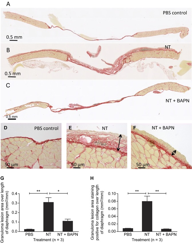
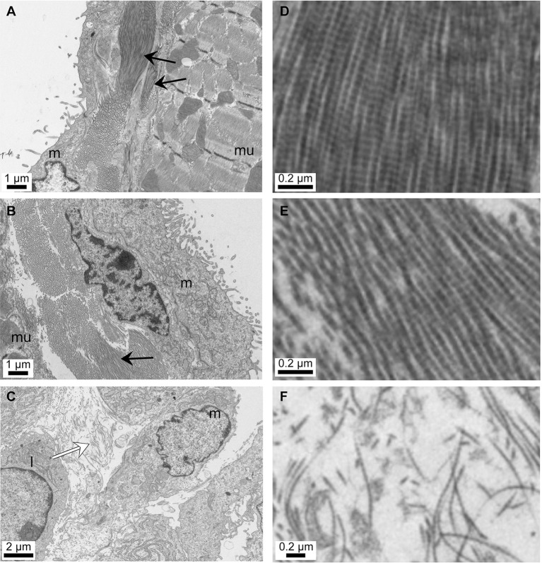
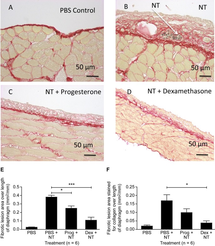
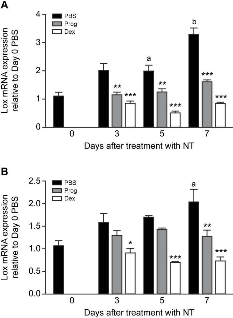
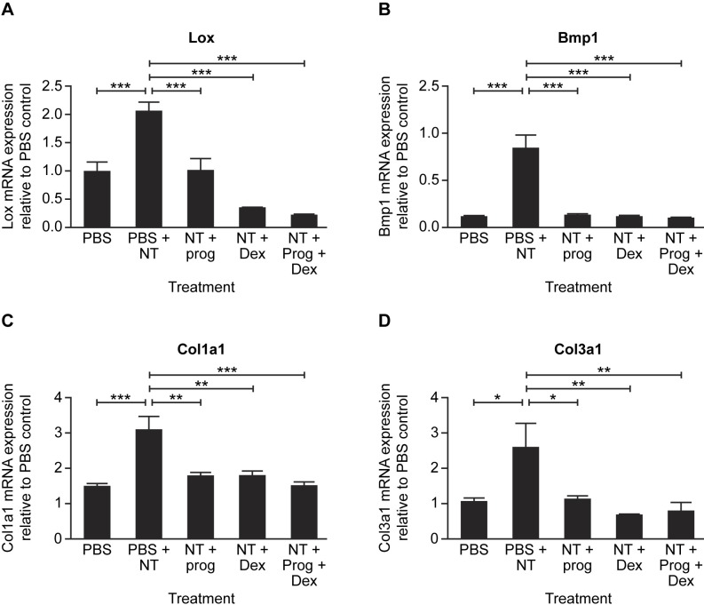
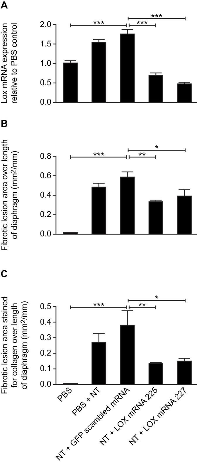
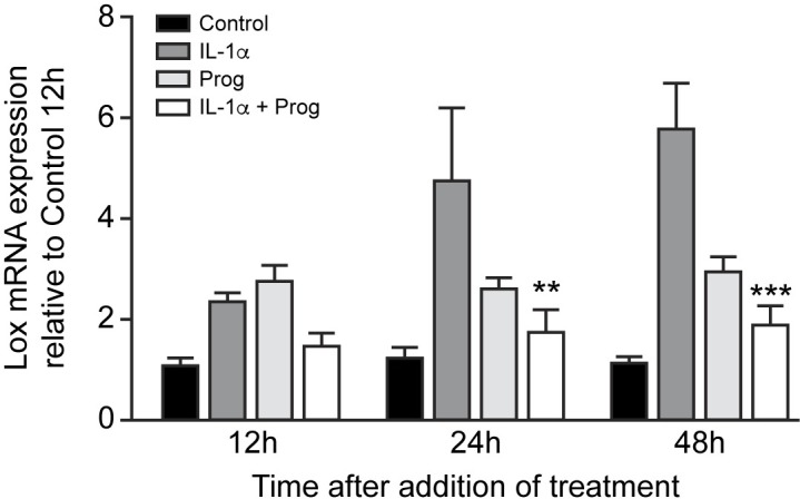
Methods: Female C57Bl/6 mice were treated intraperitoneally with multiwalled carbon nanotubes (NT) to induce fibrosis, together with chemical (ß-aminoproprionitrile-BAPN) or miRNA Lox inhibitors, progesterone or dexamethasone. Fibrotic lesions on the diaphragm, and expression of fibrosis-related genes in abdominal wall peritoneal mesothelial cells (PMC) were measured. Effects of BAPN and dexamethasone on collagen fibre alignment were observed by TEM. Isolated PMC were cultured with interleukin-1 alpha (IL-1α) and progesterone to determine effects on Lox mRNA in vitro.

Results: NT-induced fibrosis and collagen deposition on the diaphragm was ameliorated by BAPN, Lox miRNA, or steroids. BAPN and dexamethasone disrupted collagen fibres. NT increased PMC Lox, Col1a1, Col3a1 and Bmp1 mRNA, which was inhibited by steroids. Progesterone significantly inhibited IL-1α induced Lox expression by PMC in vitro.

Conclusion: Our results provide proof-of-concept that targeting peritoneal Lox could be an effective approach in ameliorating fibrosis and adhesion development.


Conflict of interest statement

Competing Interests: The authors have declared that no competing interests exist.

Figures

Fig 1
Fig 1. BAPN reduces NT-induced fibrosis on the diaphragm.
Effect of i.p. treatment with carbon nanotubes (NT, 25 μg) followed by daily injections for 7 days of the lox inhibitor ß-aminoproprionitrile (BAPN, 1 g/kg) on fibrosis and collagen deposition on the diaphragm. Picrosirius Red stained sections of control diaphragm treated with PBS (A,D), NT (B,E) and NT+BAPN (C,F). Arrows indicate the thickness of the fibrotic lesion. Red staining indicates collagen. Fibrotic granuloma lesion area (G) and extent of collagen staining within the lesion (H) were quantified. Results are expressed as mean ± SEM, n = 3. *, p<0.05; **, p<0.01. One-way ANOVA with Tukey’s multiple comparison post-hoc testing.
Fig 2
Fig 2. Electron micrographs of mouse diaphragm showing effect of NT and BAPN on sub-epithelial collagen deposition.
A,D PBS control; B,E 7 days after NT treatment; C,F 7 days after NT plus daily β-aminopropionitrile (BAPN) treatment. m: mesothelial cells, mu: muscle fibres, l: probable leukocyte. ↑ dense, ordered collagen fibre bundles, ⇧ loose, disordered collagen fibres.
Fig 3
Fig 3. Progesterone and dexamethasone reduce NT-induced fibrosis on the diaphragm.
Effect of i.p. treatment with carbon nanotubes (NT, 25 μg) followed by 7 daily injections of PBS (vehicle), progesterone (10 μg) or dexamethasone (1 μg) on fibrosis and collagen deposition on the diaphragm. Picrosirius Red stained sections of control diaphragm treated with PBS (A), NT (B), NT + progesterone (C) and NT + dexamethasone (D). Red staining indicates collagen. Fibrotic granuloma lesion area (E) and extent of collagen staining within the lesion (F) were quantified. Results are expressed as mean ± SEM, n = 6. *, p<0.05; ***, p<0.001. One-way ANOVA with Tukey’s multiple comparison post-hoc testing.
Fig 4
Fig 4. Time-dependent effects of progesterone and dexamethasone on NT-induced fibrotic gene expression in abdominal wall PMC.
Effect of i.p treatment with carbon nanotube (NT, 25 μg) followed by daily injection for up to 7 days with PBS (vehicle), progesterone (Prog, 10 μg/kg) or dexamethasone (Dex, 1 μg/kg) on Lox (A) and Col1a1 (B) mRNA expression by abdominal wall peritoneal mesothelial cells (PMC) in vivo. mRNA expression is shown relative to control (day 0 untreated animals) and is the mean ± SEM of 6–8 animals per treatment. a, p<0.05; b, p<0.01 compared with day 0 untreated animals (PBS). *, p<0.05; **, p<0.01; ***, p<0.001 compared with corresponding treatment with PBS. Two-way ANOVA with Bonferroni multiple comparison post-hoc testing.
Fig 5
Fig 5. Progesterone and dexamethasone reduce NT-induced fibrotic gene expression in abdominal wall PMC.
Effect of i.p treatment with carbon nanotube (NT, 25 μg) followed by daily injection for 7 days with PBS (vehicle), progesterone (Prog, 10 μg/kg), dexamethasone (Dex, 1 μg/kg) or Prog+Dex on lox (A), bone morphogenetic protein 1 (Bmp1) (B), Col1a1 (C) and Col3a1 (D) mRNA expression by abdominal wall peritoneal mesothelial cells in vivo. mRNA expression is shown relative to control day 0 untreated animals (PBS) and is the mean ± SEM of 8–10 animals per treatment. *, p<0.05; **, p<0.01; ***, p<0.001. One-way ANOVA with Tukey’s multiple comparison post-hoc testing.
Fig 6
Fig 6. Effect of lentiviral lox miRNA construct on carbon nanotube (NT) induced fibrosis on the abdominal wall and diaphragm.
Vehicle, scrambled miRNA and two selected Lox miRNA lentiviral constructs were injected i.p. followed 2 days later by 25 μg NT (except for the control group which received vehicle (PBS)). A), Lox mRNA in abdominal wall peritoneal mesothelial cells; B) fibrotic lesion area and C), extent of collagen staining within the lesion on the diaphragm. *, p<0.05; **, p<0.01; ***, p<0.001. One-way ANOVA with Tukey’s multiple comparison post-hoc testing.
Fig 7
Fig 7. Time dependent effects of IL-1α (2.5 ng/ml and progesterone (Prog) (1 μM) on Lox mRNA expression by abdominal wall peritoneal mesothelial cells in vitro.
mRNA expression is shown relative to control cultures at 12 h and is the mean ± SEM of 3 separate cultures using cells obtained from 6 mice. **, p<0.01; ***, p<0.01 compared to corresponding treatment with IL-1α alone. Two-way ANOVA with Bonferroni multiple comparison post-hoc testing.

References

    1. Vercellini P, Viganò P, Somigliana E, Fedele L. Endometriosis: pathogenesis and treatment. Nat Rev Endocrinol. 2014;10: 261–275. doi: 10.1038/nrendo.2013.255 - DOI - PubMed
    1. Pados G, Venetis CA, Almaloglou K, Tarlatzis BC. Prevention of intra-peritoneal adhesions in gynaecological surgery: theory and evidence. Reprod Biomed Online. 2010; 21: 290–293. doi: 10.1016/j.rbmo.2010.04.021 - DOI - PubMed
    1. Ahmad G, Mackie FL, Iles DA, O’Flynn H, Dias S, Metwally M. et al. Fluid and pharmacological agents for adhesion prevention after gynaecological surgery. Cochrane Database of Systematic Reviews. 2014; Issue 7, Art No: CD001298. - PubMed
    1. Jansen RP. Controlled clinical approaches to investigating the prevention of peritoneal adhesions. Prog Clin Biol Res. 1990;358: 177–192. - PubMed
    1. Fayez JA, Schneider PJ. Prevention of pelvic adhesion formation by different modalities of treatment. Am J Obstet Gynecol. 1987;157: 1184–1186. - PubMed

MeSH terms