Comparative Study
. 2017 Nov;38(11):1236-1248.
doi: 10.1177/1071100717723128. Epub 2017 Aug 11.

Application of High-Speed Dual Fluoroscopy to Study In Vivo Tibiotalar and Subtalar Kinematics in Patients With Chronic Ankle Instability and Asymptomatic Control Subjects During Dynamic Activities

Affiliations
Comparative Study

Application of High-Speed Dual Fluoroscopy to Study In Vivo Tibiotalar and Subtalar Kinematics in Patients With Chronic Ankle Instability and Asymptomatic Control Subjects During Dynamic Activities

Koren E Roach et al. Foot Ankle Int. 2017 Nov.

Abstract

Background: Abnormal angular and translational (ie, kinematic) motion at the tibiotalar and subtalar joints is believed to cause osteoarthritis in patients with chronic ankle instability (CAI).

Methods: In this preliminary study the investigators quantified and compared in vivo tibiotalar and subtalar kinematics in 4 patients with CAI (3 women) and 10 control subjects (5 men) using dual fluoroscopy during a balanced, single-leg heel-rise and treadmill walking at 0.5 and 1.0 m/s.

Results: During balanced heel-rise, 69%, 54%, and 66% of mean CAI tibiotalar internal rotation/external rotation (IR/ER), subtalar inversion/eversion, and subtalar IR/ER angles, respectively, were outside the 95% confidence intervals of control subjects. During 0.5-m/s gait, 50% and 60% of mean CAI tibiotalar dorsi/plantarflexion and subtalar IR/ER angles, respectively, were outside the 95% confidence intervals of control subjects. During 1.0-m/s gait, 62%, 65%, and 73% of mean CAI subtalar dorsi/plantarflexion, inversion/eversion, and IR/ER, respectively, were outside the 95% confidence intervals of control subjects. Patients with CAI exhibited less tibiotalar and subtalar translational motion during gait; no clear differences in translations were noted during balanced heel-rise.

Conclusion: Overall, the balanced heel-rise activity exposed more tibiotalar and subtalar kinematic variation between patients with CAI and control subjects. Therefore, weight-bearing activities involving large range of motion, balance, and stability may be best for studying kinematic adaptations in patients with CAI.

Clinical relevance: These preliminary results suggest that patients with CAI require more tibiotalar external rotation, subtalar eversion, and subtalar external rotation during weight-bearing stability exercises, all with less overall joint translation.

Keywords: chronic ankle instability; dual fluoroscopy; heel-rise; tibiotalar and subtalar kinematics; treadmill gait.


Figures
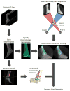
Figure 1
Figure 1
Flowchart of the methodological approach. A computed tomography (CT) scan was obtained of the subject’s ankle and foot and segmented to create three-dimensional (3-D) reconstructions of the tibia, talus and calcaneus bones. A digitally reconstructed radiograph (DRR) was created from each segmented bone. Anatomical coordinate systems were defined for the tibia, talus and calcaneus based on landmarks visible on the 3-D surfaces. Separately, dual fluoroscopy images were acquired. The dual fluoroscopy images and digitally reconstructed radiographs were used by model-based markerless tracking software to quantify the position and orientation of each bone. Bone positions and orientations were used to calculate angles and translations for the tibiotalar and subtalar joints.
Figure 2
Figure 2
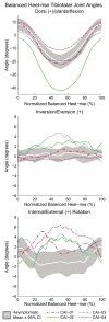
Tibiotalar dorsi (+)/plantarflexion (top), inversion/eversion (+) (middle), and internal/external (+) rotation (bottom) mean joint angles of patients with chronic ankle instability (CAI) compared to asymptomatic control subjects during a single-leg balanced heel-rise activity. Data are plotted per normalized balanced heel-rise. The joint angles of the asymptomatic control subjects are presented as the mean (white line) ± 95% confidence intervals (gray). Each colored line represents the mean joint angles of a different CAI patient.
Figure 3
Figure 3
Subtalar dorsi (+)/plantarflexion (top), inversion/eversion (+) (middle), and internal/external (+) rotation (bottom) mean joint angles of patients with chronic ankle instability (CAI) compared to asymptomatic control subjects during a single-leg balanced heel-rise activity. Data are plotted per normalized balanced heel-rise. The joint angles of the asymptomatic control subjects are presented as the mean (white line) ± 95% confidence intervals (gray). Each colored line represents the mean joint angles of a different CAI patient.
Figure 4
Figure 4
Tibiotalar dorsi (+)/plantarflexion (top), inversion/eversion (+) (middle), and internal/external (+) rotation (bottom) mean joint angles of patients with chronic ankle instability (CAI) compared to asymptomatic control subjects during 0.5 m/s (left) and 1.0 m/s (right) gait. Data are plotted per normalized stance with all subjects aligned at heelstrike (0%) and toe-off (100%). The heelstrike and toe-off portions of stance were collected as separate trials, since the movement of the treadmill caused the foot to move out of the combined field-of-view of the fluoroscopes prior to the completion of the stance phase of gait. The joint angles of the asymptomatic control subjects are presented as the mean (white line) ± 95% confidence intervals (gray). Each colored line represents the mean joint angles of a different CAI patient.
Figure 5
Figure 5
Subtalar dorsi (+)/plantarflexion (top), inversion/eversion (+) (middle), and internal/external (+) rotation (bottom) mean joint angles of patients with chronic ankle instability (CAI) compared to asymptomatic control subjects during 0.5 m/s (left) and 1.0 m/s (right) gait. Data are plotted per normalized stance with all subjects aligned at heelstrike (0%) and toe-off (100%). The heelstrike and toe-off portions of stance were collected as separate trials, since the movement of the treadmill caused the foot to move out of the combined field-of-view of the fluoroscopes prior to the completion of the stance phase of gait. The joint angles of the asymptomatic control subjects are presented as the mean (white line) ± 95% confidence intervals (gray). Each colored line represents mean joint angles of a different CAI patient.
Figure 6
Figure 6
Joint angle range of motion (ROM) values for chronic ankle instability (CAI) patients (colored symbols) plotted against the mean (black dots) ROM and 95% confidence interval (CI) (black bars) of asymptomatic controls for the tibiotalar (top) and subtalar (bottom) joints during balanced heel-rise (left), 0.5 m/s captured stance (middle), and 1.0 m/s captured stance (right). D/P = dorsi/plantarflexion; In/Ev = inversion/eversion; IR/ER = internal/external rotation.
Figure 7
Figure 7
Joint translation range of motion (ROM) values for chronic ankle instability (CAI) patients (colored symbols) plotted against the mean (black dots) ROM and 95% confidence interval (CI) (black bars) of asymptomatic controls for the tibiotalar (top) and subtalar (bottom) joints during balanced heel-rise (left), 0.5 m/s captured stance (middle), and 1.0 m/s captured stance (right). Direction of translational movement: ML = medial-lateral; AP = anterior-posterior; SI = superior-inferior.

References

    1. Bassett FH, 3rd, Gates HS, 3rd, Billys JB, Morris HB, Nikolaou PK. Talar impingement by the anteroinferior tibiofibular ligament. A cause of chronic pain in the ankle after inversion sprain. J Bone Joint Surg Am. 1990;72(1):55–59. - PubMed
    1. Bauman JM, Chang YH. High-speed X-ray video demonstrates significant skin movement errors with standard optical kinematics during rat locomotion. J Neurosci Methods. 2010;186(1):18–24. doi: 10.1016/j.jneumeth.2009.10.017. - DOI - PMC - PubMed
    1. Bey MJ, Zauel R, Brock SK, Tashman S. Validation of a new model-based tracking technique for measuring three-dimensional, in vivo glenohumeral joint kinematics. J Biomech Eng. 2006;128(4):604–609. doi: 10.1115/1.2206199. - DOI - PMC - PubMed
    1. Bischof JE, Spritzer CE, Caputo AM, et al. In vivo cartilage contact strains in patients with lateral ankle instability. J Biomech. 2010;43(13):2561–2566. doi: 10.1016/j.jbiomech.2010.05.013. - DOI - PMC - PubMed
    1. Brodsky AR, O’malley MJ, Bohne WH, Deland JA, Kennedy JG. An analysis of outcome measures following the Brostrom-Gould procedure for chronic lateral ankle instability. Foot Ankle Int. 2005;26(10):816–819. - PubMed

Publication types

LinkOut - more resources