. 2017 Aug 11;7(1):7950.
doi: 10.1038/s41598-017-08436-6.

Disrupting glutamine metabolic pathways to sensitize gemcitabine-resistant pancreatic cancer

Affiliations

Disrupting glutamine metabolic pathways to sensitize gemcitabine-resistant pancreatic cancer

Ru Chen et al. Sci Rep. .

Abstract

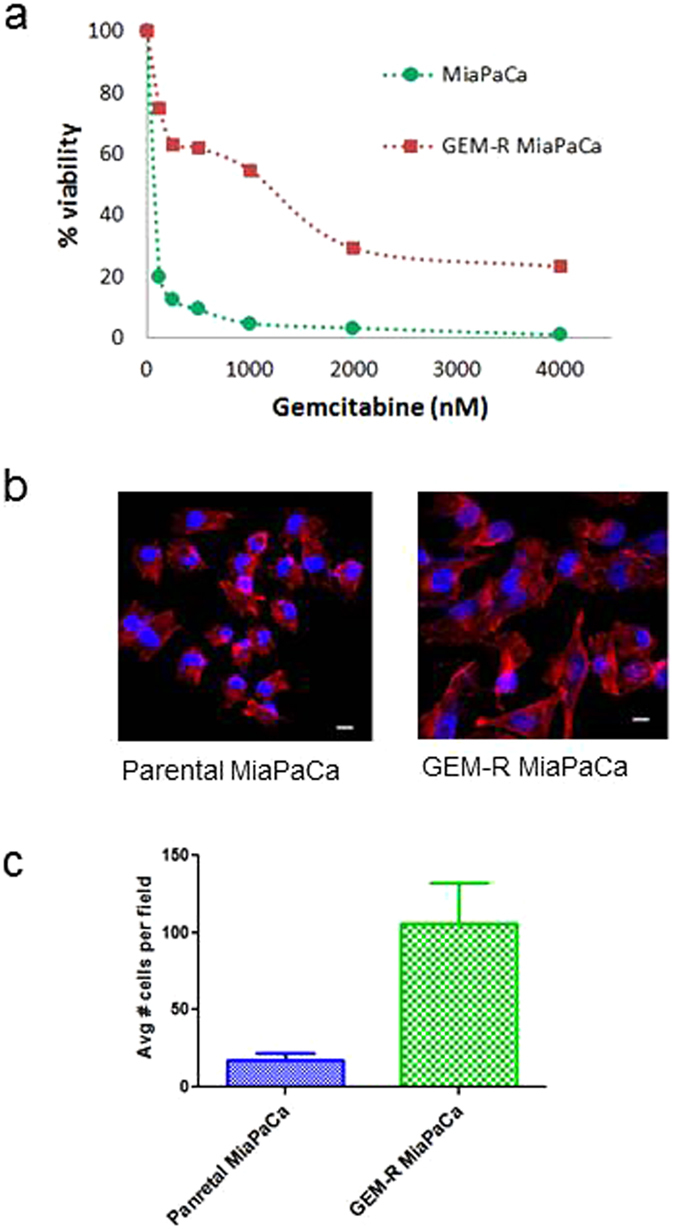
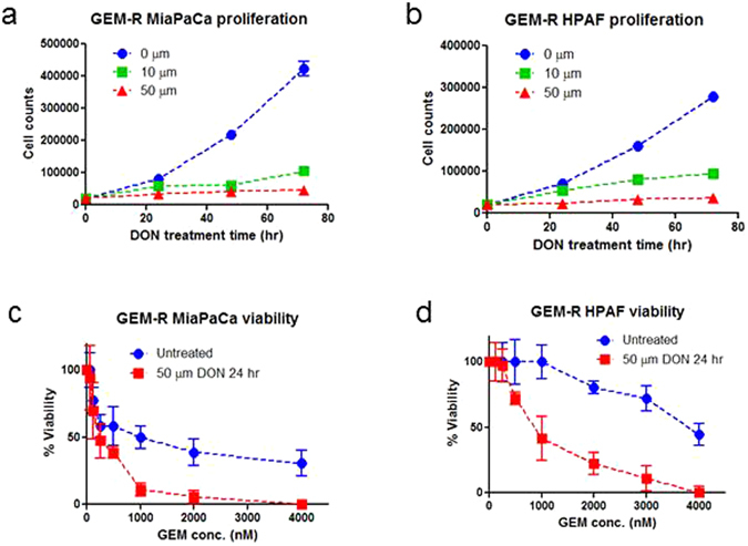
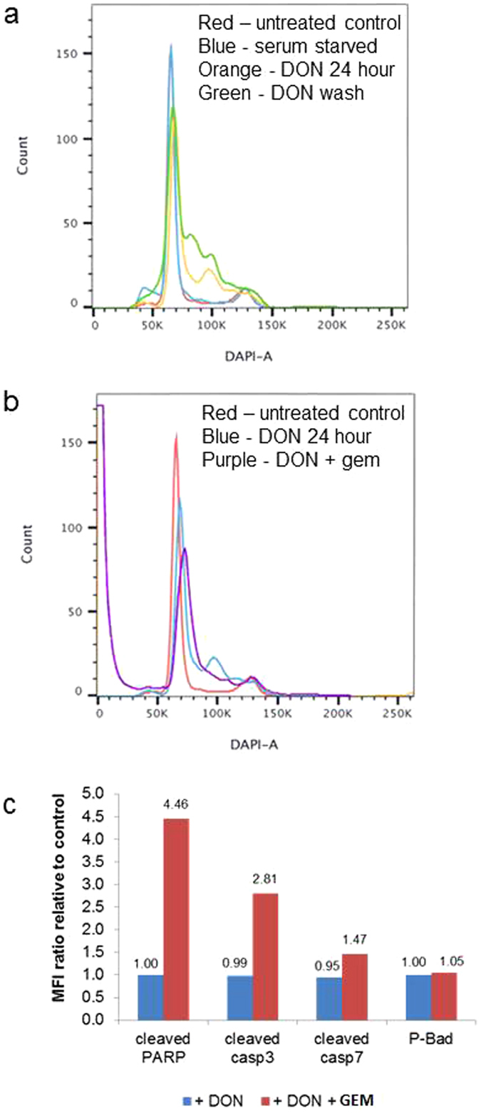
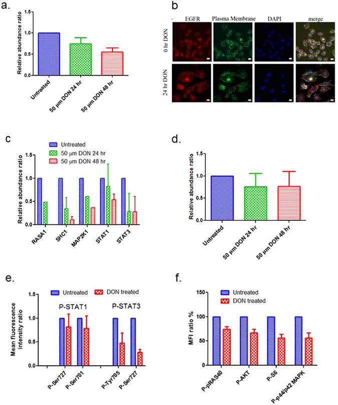
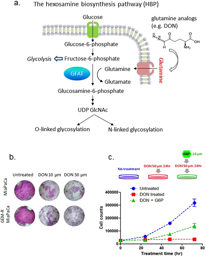
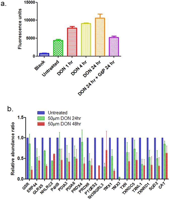
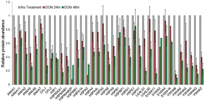
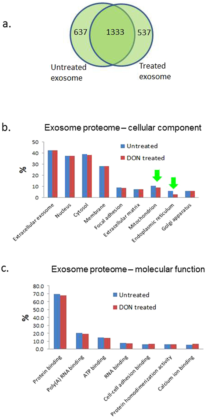
Pancreatic cancer is a lethal disease with poor prognosis. Gemcitabine has been the first line systemic treatment for pancreatic cancer. However, the rapid development of drug resistance has been a major hurdle in gemcitabine therapy leading to unsatisfactory patient outcomes. With the recent renewed understanding of glutamine metabolism involvement in drug resistance and immuno-response, we investigated the anti-tumor effect of a glutamine analog (6-diazo-5-oxo-L-norleucine) as an adjuvant treatment to sensitize chemoresistant pancreatic cancer cells. We demonstrate that disruption of glutamine metabolic pathways improves the efficacy of gemcitabine treatment. Such a disruption induces a cascade of events which impacts glycan biosynthesis through Hexosamine Biosynthesis Pathway (HBP), as well as cellular redox homeostasis, resulting in global changes in protein glycosylation, expression and functional effects. The proteome alterations induced in the resistant cancer cells and the secreted exosomes are intricately associated with the reduction in cell proliferation and the enhancement of cancer cell chemosensitivity. Proteins associated with EGFR signaling, including downstream AKT-mTOR pathways, MAPK pathway, as well as redox enzymes were downregulated in response to disruption of glutamine metabolic pathways.


Conflict of interest statement

The authors declare that they have no competing interests.

Figures

Figure 1
Figure 1
Development of GEM-R MiaPaCa PDAC cells. (a) Comparison of GEM-R MiaPaCa cells with the parental MiaPaCa in dose response to GEM treatment. (b) Cell morphological images of GEM-R MiaPaCa cells (right) and the parental MiaPaCa cells (left). (c) Cell migration assay - the number of cells migrating through the transwell was 4x higher in GEM-R MiaPaCa compared to the parental cells. The error bars represent SDs.
Figure 2
Figure 2
DON treatment of GEM-R pancreatic cancer cells significantly reduced cell proliferation and enhanced chemosensitivity. (a) Cell proliferation curves of GEM-R MiaPaCa treated with different concentrations of DON. (b) Cell proliferation curves of GEM-R HPAF II treated with different concentration of DON. (c) GEM-R MiaPaCa cells treated with 50 µm of DON for 24 hours showed significantly enhanced GEM efficacy. The IC50 value decreased 4 fold. (d) GEM-R HPAF II cells treated with 50 µm of DON for 24 hours showed significantly enhanced GEM efficacy. The IC50 value decreased 4 fold.
Figure 3
Figure 3
(a) Cell cycle analysis of GEM-R MiaPaCa with no treatment (control), incubation with 50 mM DON for 24 hours, exposure to 50 mM DON for 24 hours followed by media change and growth for 24 hours (DON washout). Note the transient G1/S phase block which was reversible following change to fresh media. (b) Cell cycle analysis of GEM-R MiaPaCa with no treatment (control), incubation with 50 mM DON for 24 hours, exposure to 50 mM DON for 24 hours followed by 72 hours treatment with GEM. Note the prominent apoptotic peak in the cells treated with DON followed by GEM (purple). (c) Multiplex bead based analysis of apoptotic markers, including cleaved PARP, cleaved caspase 3, cleaved caspase 7, and P-Bad, on GEM-R MiaPaCa with incubation with 50 mM DON for 24 hours, sequential DON and GEM treatment using untreated cells (control) as a background.
Figure 4
Figure 4
DON treatment influenced the expression and localization of EGFR associated signaling proteins. (a) The abundance changes of EGFR in GEM-R MiaPaCa cells before and after treatment with 50 µm of DON (proteomics data). (b) Immunofluorescence (IF) images of EGFR in GEM-R MiaPaCa cells before and after DON treatment. red - EGFR, green – plasma membrane, blue - DAPI. (c) Abundant changes of the proteins associated with EGFR signaling in MiaPaCa cells before and after DON treatment (proteomics data). (d) Expression changes of AKT proteins before and after DON treatment (proteomics data). (e) Changes in phospho-STAT1 and phospho-STAT3 before and after DON treatment (50 µm of DON, 24 hr) (multiplex bead based analysis). Two different phosphorylation sites (amino acids are indicated) were measured for each protein. (f) Percentage mean fluorescent intensity ratios for phospho-pRas40, phospho-Akt, phospho-S6, and phospho-p44/p42 MAPK for untreated cells (blue) and DON treated cells (red) (multiplex bead based analysis). The error bars represent SDs.
Figure 5
Figure 5
DON treatment impact glycan biosynthesis and contribute to the reduced cell proliferation. (a) Disruption of HBP pathway via glutamine analogs impacts glycan biosynthesis. (b) PAS staining of GEM-R MiaPaCa cells with no treatment, and with 10 µm and 50 µm of DON treatment for 24 hr. (c) “Rescue experiment” - Supplementation of G6P abrogated the inhibition of GEM-R MiaPaCa proliferation. The addition of G6P “rescued” the GEM-R MiaPaCa cells that were treated with DON to ~50% proliferation relative to untreated cells. Blue: untreated cells, Red: cells treated with 50 µm of DON for 24 hours, and Green: cells treated with 50 µm of DON for 24 hours followed by addition of 10 µm of G6P.
Figure 6
Figure 6
DON treatment imbalanced the redox state of GEM-R MiaPaCa cells. (a) GEM-R MiaPaCa cells treated with 50 µm of DON showed increased ROS as the incubation time increased. Addition of G6P reduced the ROS level similar to the untreated cells. (b) The expression changes of proteins involved in cell redox homeostasis before and after incubation with 50 µm of DON for 24 hr and 48 hr. The error bars represent SDs.
Figure 7
Figure 7
Abundance change of proteins previously associated with chemoresistance of pancreatic cancer, including annexin family, 14-3-3 protein family, cofilins, galectins, S100 proteins and a number of heat shock proteins. The error bars represent SDs.
Figure 8
Figure 8
Comparison of protein profiles of exosomes derived from GEM-R MiaPaCa pancreatic cancer cells with and without DON treatment. (a) Comparison of protein identification in exosomes derived from GEM-R MiaPaCa cells with and without DON treatment. (b) Comparison of cellular location of proteins identified in exosomes derived from GEM-R MiaPaCa cells with and without DON treatment. (c) Comparison of molecular function of proteins identified in exosomes derived from GEM-R MiaPaCa cells with and without DON treatment.

References

    1. Siegel R, et al. Cancer treatment and survivorship statistics, 2012. CA Cancer J. Clin. 2012;62:220–241. doi: 10.3322/caac.21149. - DOI - PubMed
    1. Vincent A, Herman J, Schulick R, Hruban RH, Goggins M. Pancreatic cancer. Lancet. 2011;378:607–620. doi: 10.1016/S0140-6736(10)62307-0. - DOI - PMC - PubMed
    1. Paez D, Labonte MJ, Lenz HJ. Pancreatic cancer: medical management (novel chemotherapeutics) Gastroenterol. Clin. North Am. 2012;41:189–209. doi: 10.1016/j.gtc.2011.12.004. - DOI - PubMed
    1. Hung SW, Mody HR, Govindarajan R. Overcoming nucleoside analog chemoresistance of pancreatic cancer: a therapeutic challenge. Cancer Lett. 2012;320:138–149. doi: 10.1016/j.canlet.2012.03.007. - DOI - PMC - PubMed
    1. Pavlova NN, Thompson CB. The Emerging Hallmarks of Cancer Metabolism. Cell Metab. 2016;23:27–47. doi: 10.1016/j.cmet.2015.12.006. - DOI - PMC - PubMed

Publication types

MeSH terms