

Effect of X-ray irradiation on hepatocarcinoma cells and erythrocytes in salvaged blood
- PMID: 28801583
- PMCID: PMC5554194
- DOI: 10.1038/s41598-017-08405-z
Effect of X-ray irradiation on hepatocarcinoma cells and erythrocytes in salvaged blood
Abstract
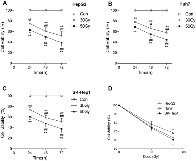
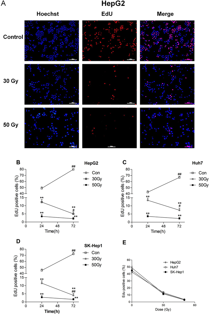
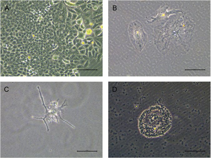
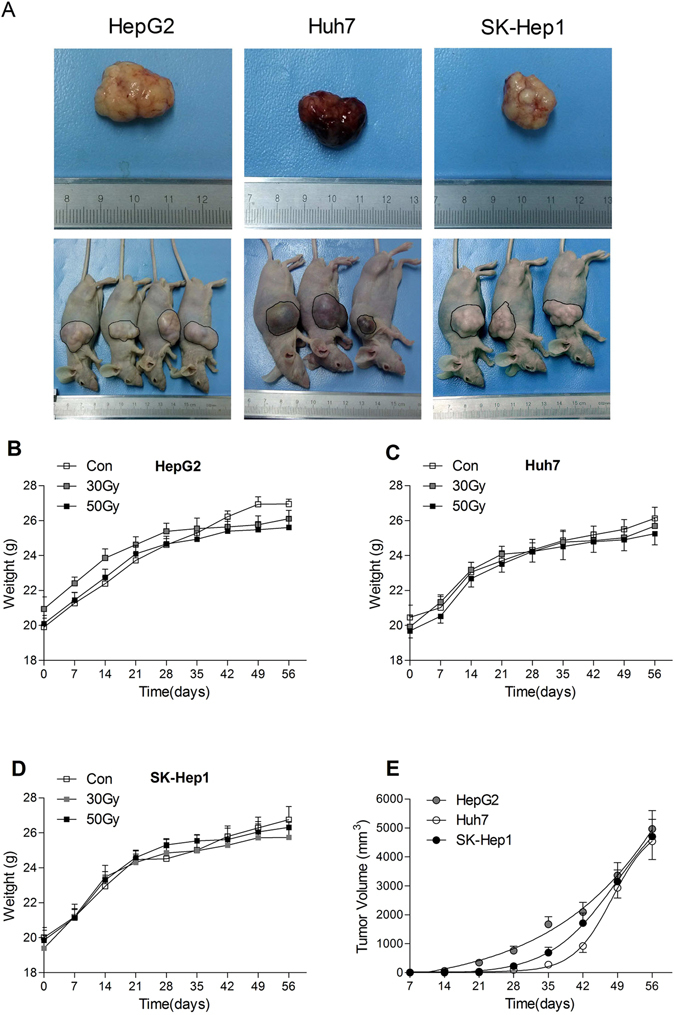
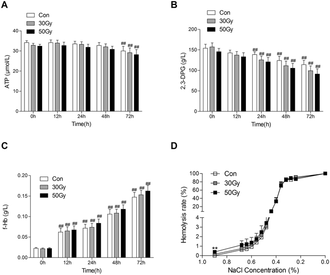
The broad clinical acceptance of intraoperative blood salvage and its applications in cancer surgery remain controversial. Until now, a method that can safely eliminate cancer cells while preserving erythrocytes does not exist. Here, we investigated whether X-ray generated from linear accelerator irradiation at a certain dose can kill hepatocarcinoma cells while preserving erythrocytes. HepG2, SK-Hep1 or Huh7 cells were mixed into the aliquots of erythrocytes obtained from healthy volunteers. After the mixed cells were exposed to 30 Gy and 50 Gy X-rays irradiation, the viability, clonogenicity, DNA synthesis and tumorigenicity of the tumor cells were determined by the MTT assay, plate colony formation, 5-ethynyl-2'-deoxyuridine incorporation, and subcutaneous xenograft implantation into immunocompromised mice. The ATP, 2,3-DPG, free Hb, osmotic fragility, blood gas variables in erythrocytes and morphology of erythrocytes at 0 h, 12 h, 24 h, 48 h, 72 h after irradiation were analyzed. X-ray irradiation at 30 Gy effectively inhibited the viability, proliferation, and tumorigenicity of HepG2, SK-Hep1 and Huh7 cells without noticeably damaging the ability of oxygen-carrying, membrane integrity and morphology of erythrocytes. Theses results suggest that X-ray at 30 Gy irradiation might be safe to eliminate hepatocarcinoma cells while preserving erythrocytes in salvaged blood.
Conflict of interest statement
The authors declare that they have no competing interests.
Figures
References
Publication types
MeSH terms
LinkOut - more resources
Full Text Sources
Other Literature Sources
Medical
